
Abstract
Objective:
Annular elastolytic giant cell granuloma (AEGCG) is a rare granulomatous disease characterized by annular plaques. In this study, we aimed to describe the clinical and pathological features of the patients diagnosed with AEGCG.
Methods:
The demographic, clinical and pathological features of patients who diagnosed with AEGCG were recorded retrospectively.
Results:
Ten patients with AEGCG included in the study (nine females and one male). The mean age of the patients was 60±9.53 years. The mean duration of disease was 24.2±36.30 months. On dermatologic examination, multiple, well-demarcated, elevated borders and central atrophic erythematous annular plaques were seen in all patients. In the most of the patients (90%) lesions were on the sun-exposed regions. Six of the patients had accompanying diseases. Histopathologic examination of the punch biopsies revealed foreign body type multinucleated giant cells and lymphocytic cell infiltration in the dermis. There were intracellular elastic fiber fragments as sign of elastophagocytosis in the giant cells.
Conclusion:
AEGCG is a rare granulomatous disease which can accompany various diseases. There is debate on the terminology, classification and pathogenesis. Further studies are required to elucidate the unknowns.
Keywords: Annular elastolytic giant cell granuloma, elastophagocytosis, granulomatous diseases, elastolysis, giant cell, granuloma annulare
Introduction
Annular elastolytic giant cell granuloma (AEGCG) is a rare granulomatous disease characterized by annular plaques with elevated borders and central atrophy. It was first defined by Hanke et al. (1) in 1979. He described annular patches with the histopathological appearance of many multinucleated giant cells, total lysis of elastic tissue, and the absence of necrobiosis, mucin or lipids (1,2).
The exact pathogenesis of AEGCG is unknown. It is thought that exposure to the sun, heat or other factors change the antigenicity of the elastic fibers and causes cellular immune reactions. Elastophagocytosis and granuloma formation may reflect the inflammatory reaction against elastic fibers (2).
In our study, we aimed to describe the clinical and histopathologic features of AEGCG based upon 10 patients diagnosed with AEGCG and reviewed the literature.
Methods
We evaluated 10 patients histopathologically diagnosed with AEGCG who attended to our dermatology department between 2006 and 2017. Demographic (age, sex), clinical (duration, localization, accompanying disease and treatment) and pathological features of the cases were noted retrospectively from the patients’ files.
Results
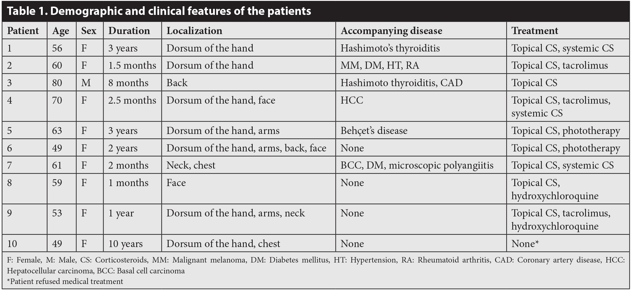
The study included 10 patients diagnosed with AEGCG (9 females and 1 male). Mean age of patients was 60±9.53 years. Demographic and clinical features of the patients are shown in .
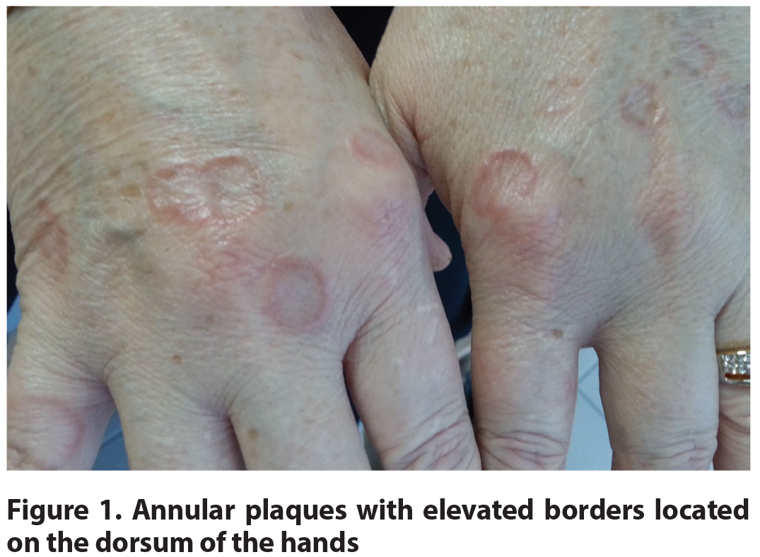
On dermatologic examination, all of the patients had multiple, well-demarcated, erythematous annular and serpiginous plaques with elevated borders and central atrophy. Patients revealed that lesions began as papules, then the papules expanded centrifugally to annular plaques (). Most of the patients (90%) had lesions on sun-exposed regions such as dorsum of hands, arms, face, neck and chest. Only 1 patient’s lesions located on the back. Mucosa, scalp and nail examinations were normal. None of the patients had complaints such as itching, burning or pain.
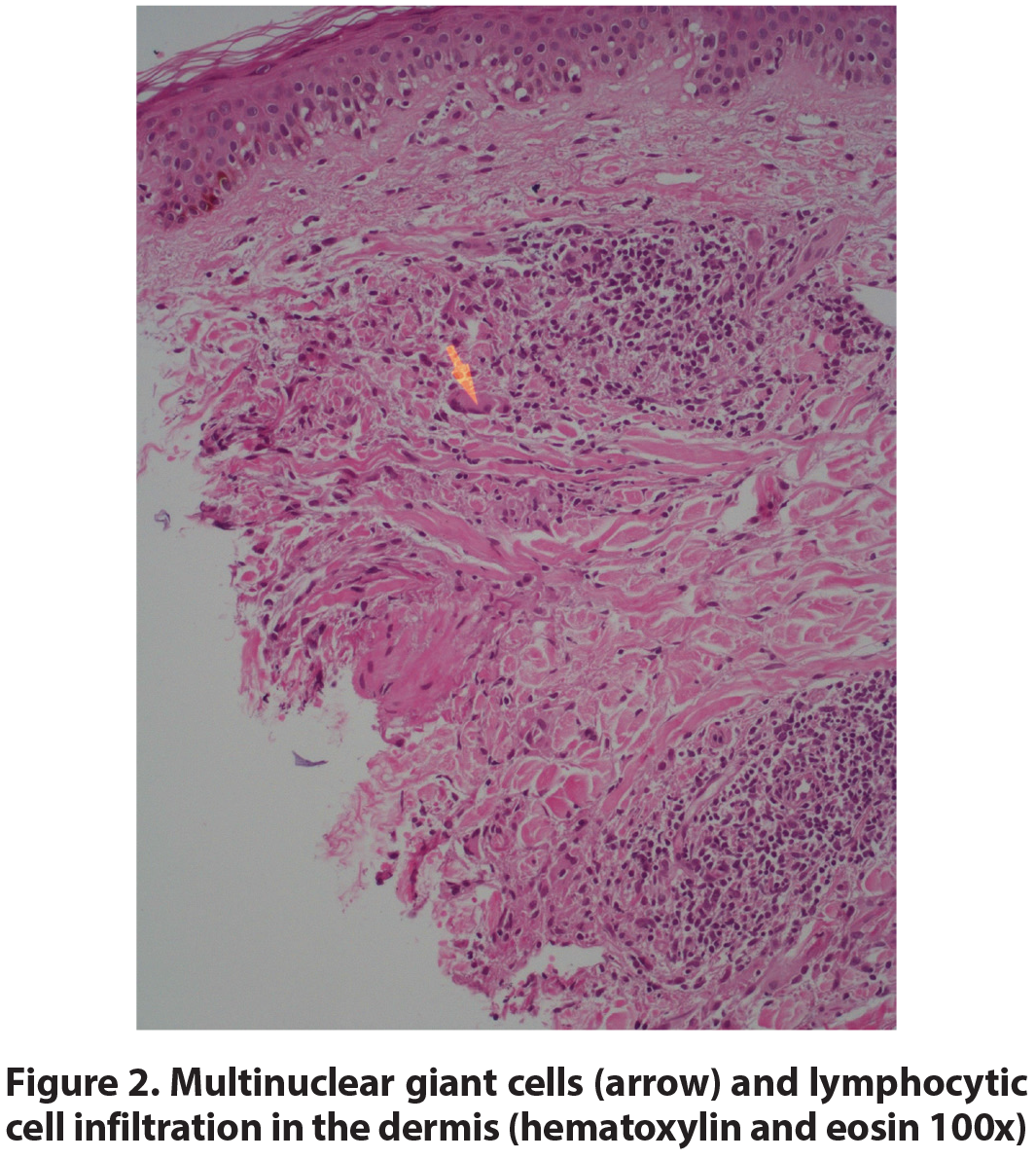
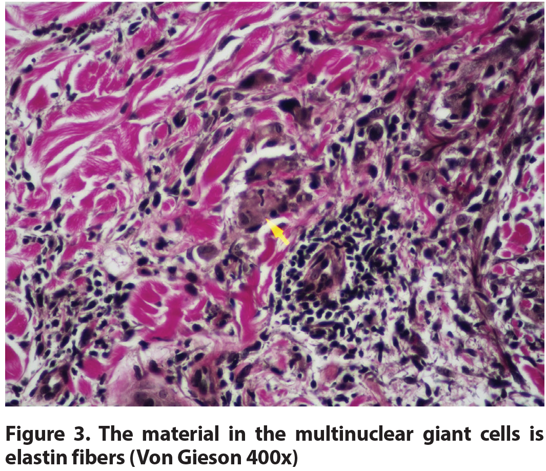
One 4 mm punch biopsy was taken from each patient with the pre-diagnoses of granuloma annulare, AEGCG, sarcoidosis, erythema annulare centrifugum, pityriasis rosea and subacute lupus erythematosus. Histopathologic examination of the punch biopsies revealed foreign body type multinuclear giant cells and lymphocytic cell infiltration in the dermis. A palisading arrangement of the histiocytes was not a feature. Giant cells had intracellular elastin fiber fragments as sign of elastophagocytosis ( and ). Epidermis was atrophic in 2 and normal in 8 of the biopsies. A mild lymphohistiocytic infiltrate was present around small vessels of the upper and mid dermis. Of the 6 biopsies there was also variable component of plasma cells and eosinophils. In the surrounding dermis, typical solar elastosis was not a consistent feature. The dermal collagen is relatively normal. In differential diagnosis, granuloma annulare and necrobiosis lipoidica were considered. Granuloma annulare was excluded due to absence of elastophagocytosis and mucin deposition. As well as the absence of necrobiosis and palisading granulomas necrobiosis lipoidica was excluded. Fungal and mycobacterial infections were excluded with periodic acid schiff and Ziehl-Neelsen stains.
All of the patients revealed progressive disease with sudden onset. The mean disease duration was 24.2±36.30 months, varying between 1 month to 10 years. Six of the patients had accompanying diseases as follows: Hashimoto’s thyroiditis in 2 patients, diabetes mellitus in 2 patients, hypertension in 1 patient, coronary artery disease in 1 patient, microscopic polyangiitis in 1 patient, Behçet’s disease in 1 patient and the history of malignancy in 3 patients (hepatocellular carcinoma, basal cell carcinoma and malignant melanoma; 1 patient for each disease). Although cancer patients were in remission period with no signs of malignancy; we scanned all patients to rule out the malignancies with detailed physical examination (including lymph node examination), complete blood count, liver function tests, renal function tests, serum lactic dehydrogenase, urinalysis and chest X-ray examination. They were all within normal limits.
Discussion
AEGCG lesions are clinically characterized by papules or annular plaques with erythematous-elevated borders and an atrophic center located predominantly on sun-exposed areas. Most of the patients are middle-aged, white women. The lesions are generally asymptomatic and heal without scarring (1,3-5).
Annular lesions are challenging for dermatologist in clinical practice. Clinical differential diagnosis of annular lesions includes erythema annulare centrifugum, granuloma annulare, lichen planus, sarcoidosis, necrobiosis lipoidica, nummular eczema, leishmaniasis, syphilis, tinea corporis, leprosy, granuloma multiforme and AEGCG (4,6). Histopathological examination is frequently required for differential diagnosis.
AEGCG is a member of the elastolytic granulomas group; the others in this group are actinic granuloma, Miesher’s granuloma and atypical necrobiosis lipoidica of the face and scalp. There is a debate on the classification. Some authors use the term “annular elastolytic giant cell granuloma” to define the all cases of annular plaques with elastophagocytosis and multinucleated giant cells histopathologically. However, cases with severe solar elastosis is generally differentiated from AEGCG and called as actinic granuloma (7).
Granulomatous infiltrate with lymphocytes, histiocytes and multinucleated giant cells in the dermis, degradation of elastic fibers and elastophagocytosis by giant cells are the major histopathologic findings of AEGCG (8).
Histopathologically, the main differential diagnoses of AEGCG are granuloma annulare and necrobiosis lipoidica. Some authors concluded that this discrimination was artificial. However, necrobiosis and increased dermal mucin is absent in AEGCG unlike granuloma annulare and necrobiosis lipoidica. Elastolysis may be seen in granuloma annulare, but the complete loss of elastic fibers in the central area is characteristic of AEGCG (4,8,9). Furthermore, a hybrid pattern has been described, indicating an overlap between AEGCG and granuloma annulare (10). Consequently, histopathology of the our cases were consistent with the literature by the presence of the foreign body type multinuclear giant cells, lymphocytic cell infiltration, elastophagocytosis and the absence of solar elastosis, necrobiosis and mucin deposition.
Arora et al. (6) presented 10 cases of AEGCG. Most of their patients were in the sixth to the seventh decade. Similarly, mean age of our patients was 60±9.53 years. The female/male ratio was 1.2/1; whereas 9/1 in our study. There was only one patient associated with hypothyroidism in their study; but 80% of our patients had at least one accompanying diseases (6).
In the literature, AEGCG cases associated with malignancies such as prostate carcinoma, acute myelogenous leukaemia, adult T-cell leukaemia, primary cutaneous T-cell lymphoma and squamous cell carcinoma of the tonsils have been reported (3,11-13). AEGCG may emerge as a systemic immunologic host defense against the tumor antigens (14). Paraneoplastic feature of AEGCG can be supported by disappearance of AEGCG with the treatment of underlying malignancy and concomitant recurrence of both the malignancy and the AEGCG. Conversely, Asahina et al. (11) reported a case of AEGCG associated with prostate carcinoma that fail to improve despite removal of the tumor. Thus, they suggested that this association might be only coincidental. They also showed metalloproteinase-12 upregulation in AEGCG lesions and put forward the role of matrix metalloproteinase in degradation of elastic fibers (11). In our study, the history of malignancy was detected in 3 patients (hepatocellular carcinoma, basal cell carcinoma and malignant melanoma; 1 patient for each disease). Patients were in remission period and we excluded recurrence with physical examination and laboratory tests. Our study is the first report of the association between AEGCG and hepatocellular carcinoma, basal cell carcinoma and malignant melanoma. AEGCG can develop due to immunologic host defense against the tumor antigens, or it can be merely coincidental. Consequently, dermatologist should keep in mind the paraneoplastic nature of the AEGCG and scan patients to rule out the malignancies.
AEGCG can also be accompanied by systemic and dermatologic disorders such as diabetes mellitus, Hashimato’s thyroiditis, stroke, temporal arteritis and vitiligo (5,8,15-17). In our study diabetes mellitus, Hashimato’s thyroiditis, hypertension, coronary artery disease, microscopic polyangiitis, Behçet’s disease were found to be accompanying diseases to AEGCG. Aso et al. (15) noticed that nearly 37% of Japanese AEGCG patients were found to have diabetes mellitus. This ratio was 20% in our study. Diabetes mellitus may induce AEGCG development probably by damaging the elastic fibers (15). Moreover, AEGCG cases associated with autoimmune disorders such as Hashimato’s thyroiditis and vitiligo may suggest the role of autoimmunity in the pathogenesis of AEGCG (8,16). Two of our patients had vasculitis (microscopic polyangiitis and Behçet’s disease). Similarly, Shoimer and Wismer (5) reported a case of AEGCG associated with vasculitis (giant cell arteritis). Authors concluded that both of the diseases were similar in terms of histopathology and pathogenesis. These two diseases developed as inflammatory response involving a granulomatous reaction to elastin fibers. Besides, histopathologically they showed granulomatous infiltration, giant cells and loss of elastic fibers or internal elastic lamina (5).
Local immune changes have also been proposed to trigger the development of AEGCG. Watabe and Akasaka (16) presented a case of AEGCG occurred on vitiligo lesions. They suggested that damaged elastic fibers in the vitiligo lesions may cause lymphocyte and macrophage accumulation, elastophagocytosis and granuloma formation (16). In another case of AEGCG developed on the contralateral extremity to cerebral ischemic stroke, authors thought that ischemia damaged motor and sensory nerves and might cause release of neuropeptides and finally induced local immunologic destabilization (17). Furthermore, local trauma was also blamed in the development of AEGCG (18,19).
Although AEGCG has a chronic course and the treatment is usually unsatisfactory; patients with spontaneous remission have also been described (1,20). There is no standard therapy and the data related with treatment options based on anecdotal case reports. In the literature, cases treated with topical, intralesional or systemic corticosteroids, hydroxychloroquine, clofazimine, cyclosporine, dapsone, methotrexate, phototherapy (psoralen plus ultraviolet A therapy and narrowband ultraviolet B therapy), retinoids, fumaric acid esters, tranilast, minocycline, cryotherapy and topical calcineurin inhibitors have been reported (2,21,22).
Study Limitations
Our study is retrospective; so it can be considered as a limitation of the study.
Conclusion
AEGCG is a granulomatous disease that can be associated with malignancies and various disorders. Although our study is retrospective; report of 10 cases is precious for this rare disease. There is a debate on the terminology and classification of AEGCG. Besides, the exact pathogenesis is still unknown. More case reports and further studies are warranted to elucidate the associations and the mysteries.
Footnotes
Ethics
Ethics Committee Approval: It wasn’t taken because this study is retrospective.
Informed Consent: It wasn’t taken because this study is retrospective.
Peer-review: Internally peer-reviewed.
Authorship Contributions
Surgical and Medical Practices: H.K.E., D.A., Concept: H.K.E., Z.N.S., Design: H.K.E., D.A., Z.N.S., Data Collection or Processing: H.K.E., D.A., E.Y., Analysis or Interpretation: H.K.E., D.A., E.A., E.Y., Z.N.S., Literature Search: H.K.E., D.A., Yazan: H.K.E.
Conflict of Interest: No conflict of interest was declared by the authors.
Financial Disclosure: The authors declared that this study received no financial support.