
Abstract
Eosinophilic cellulitis (EC), also known as Wells syndrome, presents as sudden fever, erythematous and edematous, pruritic plaques, and/or vesiculobullous lesions, and is exceptionally rare in infants. We report a case of a 7-month-old female with bilateral infantile EC resembling scleredema. The condition was characterized by acute fever, edema, and erythema from the wrists to the elbows. Histopathological examination showed spongiosis, intense inflammatory infiltration, numerous eosinophils, and collagen degeneration (flame figures), confirming EC. Treatment with systemic steroids and topical creams resulted in rapid resolution of lesions within a week, with no recurrence during a 1-year follow-up.
Keywords: Eosinophilic cellulitis, inflammatory, infantile
INTRODUCTION
Eosinophilic cellulitis (EC), also known as Wells syndrome, is an uncommon inflammatory dermatitis characterized by various clinical presentations that are often marked by a sudden onset of pruritic erythematous tender plaques. Pediatric EC is already recognized as a rare condition, and its onset in infants is exceptional. While the predominant clinical form is characterized by erythematous plaques, rare presentations include vesicle, bulla, and nodule formation. Patients with EC may experience localized symptoms such as burning and itching. In more severe cases, systemic symptoms like fever, lymphadenopathy, and arthralgia may also be present.
The localization of lesions on the extremities, presenting as erythematous plaques, can mimic infectious cellulitis, scleredema, or contact dermatitis, posing diagnostic challenges. The etiology of EC remains frequently unknown, with reported triggers including infections, tattooing, arthropod bites, and vaccinations., However, approximately half of pediatric cases lack identifiable triggers. Identifying and addressing the underlying causes is essential for preventing recurrence.
Herein, we present a case of rapid-onset EC mimicking scleredema in a 7-month-old infant and review the literature on infantile EC cases.
CASE REPORT
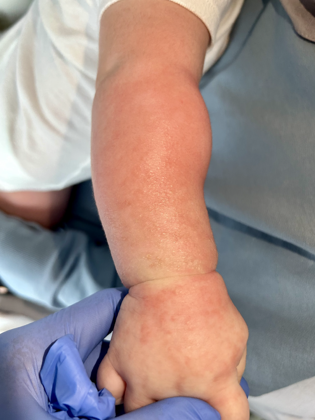
A 7-month-old female patient was admitted to the emergency department with complaints of sudden-onset fever, bilateral edema, stiffness, and redness extending from the wrists to the elbows. In the emergency room, the fever responded to paracetamol treatment, and it recurred every 4 hours. The patient was hospitalized due to recurrent fever and poor general condition. The patient’s medical history was unremarkable; she was an otherwise healthy baby delivered at full term via elective cesarean section. Her previous laboratory evaluations noted eosinophilia and a history of febrile infections, two of which required the administration of systemic antibiotics when she was four and five months old.
Further investigation into the patient’s vaccination history revealed that she had received the third dose of the hepatitis B and diphtheria, tetanus, acellular pertussis - inactivated poliovirus - haemophilus influenzae type B vaccines, as well as the first dose of the oral poliovirus vaccine, 5 days prior to admission. The patient had no history of rash following previous vaccination.
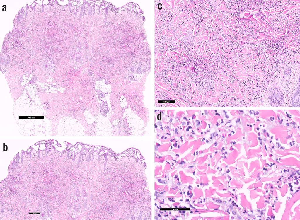
Dermatological examination revealed bilateral firmness, erythematous and edematous papules, and plaques extending from the wrists to the elbows (). A 3 mm punch biopsy was performed. The lesion showed prominent spongiosis in the epidermis, multiple spongiotic microvesicles containing eosinophils, resulting in a “Swiss cheese” appearance, intense inflammatory infiltration rich in eosinophils extending from the papillary dermis up until the mid-deep dermis, and foci of collagen degeneration, which can be described as “flame figures” ().
Complete blood count revealed leukocytosis [17.5 (5.5-17 K/uL)], mild eosinophilia [1.3 (0-1.1 K/uL)], and elevated C-reactive protein [8.1 mg/L (<5 mg/L)]. Blood cultures showed no growth. Infection serology negative for influenza A, influenza B, respiratory syncytial virus, adenovirus, and severe acute respiratory syndrome-coronavirus-2.
The patient was started on intravenous methylprednisolone at a dose of 1.6 mg/kg per day, along with topical fusidic acid and hydrocortisone acetate cream. By the second day of treatment, there was notable reduction in erythema and stiffness of the lesions, with complete resolution within a week. Systemic steroids were discontinued on the sixth day. In the 1-year follow-up period, the patient did not experience any recurrence, even after reintroducing the previous vaccinations. Caregivers have given written consent for publication.
DISCUSSION
EC, or Wells syndrome, was described in four patients by Wells as recurrent granulomatous dermatitis with eosinophilia in 1971.
Clinically, the majority of EC cases are typically observed in adults without sex predilection, with sudden onset of tender, erythematous, edematous, and well-circumscribed plaques on the extremities. Caputo et al. described seven clinical variants of EC: Classic plaque-type variant, annular granuloma-like, urticaria-like, bullous, papulonodular, papulovesicular, and fixed drug eruption-like. They also found that the classic plaque type is the most common clinical form in adults, and the annular granuloma-like form is more common in the pediatric population. Around the lesions, prodromal symptoms, such as burning and pruritus, may occur, and some patients may also experience systemic symptoms, such as fever, arthralgia, and lymphadenopathy., , 8
EC is a rare entity, and it is also rare in the pediatric population. To our knowledge, 17 cases of infantile EC under the age of 2 years have been reported to date ()., , , 9-20
Table 1
Table 1. Eosinophilic cellulitis in infancy: report of a case and literature review | ||||||||
|---|---|---|---|---|---|---|---|---|
Author | Case | Gender | Location-clinical form | Trigger | Lab abnormalities | Treatment | Recurrence | Follow-up |
Afsahi and Kassabian9 | 17-mo | Boy | Bilateral multiple tender, fluctuating, and indurated plaques on the palms, soles, and dorsum of the foot | NR | Eosinophilia (14%) | Systemic prednisone | - | 3 weeks |
Barreiros et al.10 | 18-mo | Girl | Bilateral, solitary well-demarcated, erythematous plaques on the legs | Parvovirus B19 infection | Normal | Spontaneous remission | - | 2 weeks |
Caputo et al.7 | 1-yo | Girl | Bilateral multiple papulovesicular lesions in the lower extremities | NR | NR | Topical corticosteroid, Systemic betamethasone | + | 2 years |
4-mo | Boy | Papulonodular solitary lesion on the face | NR | Eosinophilia (13%) | Systemic betamethasone | + | 3 years | |
Garty et al.11 | Newborn | Girl | Multiple subcutaneous nodules in the scalp and trunk Multiple bilateral erythematous vesicular and pustular lesions on the trunk, abdomen, inguinal regions, and wrists Bilateral multiple submandibular, axillary, and inguinal lymph nodes Hepatosplenomegaly | NR | Leukocytosis (15,000/mm3) Eosinophilia (21%) Increased erythrocyte sedimentation rate (30 mm/h) Anemia (10.5 g/dL) | Treatment with antibiotic ointments, antifungal medications, or steroids was ineffective | + | 3 years |
Gilliam et al.5 | 1-yo | Girl | Bilateral multiple erythematous, edematous, and bullous plaques on the arms and lower extremities | NR | Leukocytosis (30x109 cells per L) Eosinophilia (48%) | Combination of systemic plus topical steroids | - | 1 year |
Kamani and Lipsitz3 | 7-wk boy | Boy | Multiple bilateral erythematous plaques on the neck and shoulder Unilateral lymph nodes | NR | Leukocytosis (22,600 per mm3) Eosinophilia (16%) Increased erythrocyte sedimentation rate (60 mm/h) | Systemic prednisone | + | 2 years |
3-wk | Boy | Unilateral solitary erythematous plaque in the right thigh | NR | Leukocytosis (29,000 per mm3) Eosinophilia (32%) | Systemic prednisone | + | 6 months | |
Kuwahara et al.12 | Newborn | Girl | Unilateral firm hyperpigmented solitary plaque on the wrist | NR | NR | Spontaneous remission | - | 2 years |
Lindskov et al.13 | 20-mo | Boy | Bilateral multiple herpetiform papulovesicular lesions in all four extremities and the face | NR | Leukocytosis (20,000 per mm3) Eosinophilia (12.5%) Slight anemia Slightly elevated IgE levels | Topical antiseptics | + | 2 years |
Makni et al.14 | 14-mo | Boy | Generalized multiple erythematous papulovesicular lesions on the face, trunk, and all four extremities Multiple bilateral brownish nodular lesions on the thigh and back of the foot | NR | Leukocytosis (12,160 per mm3) Eosinophilia (10.2%) | Topical corticosteroids | - | NR |
Moon et al.15 | 5-mo | Girl | Unilateral reddish, annular plaque on the trunk | Insect bite | Normal | Topical hydrocortisone ointment | - | NR |
Moossavi and Mehregan16 | 21-mo | Girl | Bilateral multiple tense blisters on an erythematous base of the arms | NR | Normal | Oral prednisone Triamcinolone 0.1% cream | - | 1 year |
Shimshak et al.17 | 13-mo | Girl | Generalized multiple pink papules and erythematous plaques on the trunk and extremities | Varicella vaccine | Normal | Oral cetirizine Topical corticosteroid | NR | NR |
Simpson et al.18 | 22-mo | Boy | Urticarial patches on the back Bilateral erythematous, edematous papulovesicular lesion on the dorsum of the hand, ankles, and feet | Influenza vaccine | Eosinophilia (1.4x10³/µL) | Chlorpheniramine Paracetamol | + | 1 year |
Weiss et al.19 | 18-mo | Girl | Multiple bilateral erythematous papules and plaques on the buttocks | NR | Elevated eosinophilic cationic protein (85.5µ) Elevated serum IgE (22.0 kIU/L) | Topical clobetasol propionate | - | 9 months |
Wood et al.20 | 18-mo | Boy | Bilateral multiple infiltrated annular plaques on the legs | NR | Eosinophilia | NR | NR | NR |
Current case | 7-mo | Girl | Bilateral multiple erythematous, edematous papules and plaques on the arms | NR | NR | Systemic steroid Topical steroids plus antibiotics | - | 1 year |
NR: Not reported, wk: Week, mo: Month, yo: Year | ||||||||
The age range of infantile cases is 0-22 months. There are 10 girls and 8 boys, including our case. Despite the various anatomical regions affected, the extremities are the most commonly involved body parts. Three cases were triggered by vaccination and one by insect bite. Treatment included systemic corticosteroids in eight cases and topical corticosteroids in an equal number, both leading to rapid responses. Except for five cases, no recurrences were observed during follow-up. Triggering factors include bacterial and viral infections, arthropod bites, drugs, vaccinations, and malignancies. While vaccinations are the most frequently reported triggers, many pediatric cases have no identifiable cause., , , 18 In our case, although lesions appeared following vaccination, in the absence of recurrence following a subsequent dose, vaccination cannot be incriminated beyond reasonable doubt as a trigger factor. More than half of patients have transient blood eosinophilia in laboratory analysis in EC (11/18). The simultaneous presence of eosinophilia in both tissue and peripheral blood is a common finding in EC.21
The proposed mechanism suggests an external trigger that leads to elevated levels of circulating interleukin-2 (IL-2), IL-5, and eosinophil cationic protein, contributing to the activation of CD4+ T-cells and eosinophils.22 Similarly, in the literature, some studies found an increase in IL-5 and eosinophilic cationic protein in peripheral blood during the active phase of WS.23
Histopathological features of EC are typically characterized by dermal edema, dermal infiltration of histiocytes and eosinophils, and eosinophil granules surrounding collagen fibers, which are described as “flame figures”.Although the flame figure is a valuable clue to EC, it is not pathognomonic. Other diseases where flame figures may be observed include eczema, arthropod bite, severe prurigo, pemphigoid, and its variants.8
The clinical course of EC is generally benign and self-limiting, and lesions may regress spontaneously without scarring.The essentials of treatment are avoiding triggers and treating the underlying causes. Treatment strategies for EC typically include midpotency topical corticosteroids, either alone or in conjunction with systemic corticosteroids. Notably, the literature also reports the use of alternative medications, such as cyclosporine, dapsone, antimalarial drugs, and azathioprine, in certain cases series.24
Diagnosis of EC can be quite challenging for physicians because it requires careful evaluation of the patient’s medical history, including medications and vaccinations. It is essential to distinguish EC from bacterial cellulitis, contact dermatitis, granuloma annulare, urticaria, and allergic drug eruptions, which are included in the differential diagnosis., 25 Another differential diagnosis to consider is eosinophilic annular erythema (EAE). EAE is often considered a variant of Wells syndrome in the literature; however, there are notable clinical and histopathological differences between these two conditions.26 Clinicopathologic correlation plays a crucial role in differentiating EC from the abovementioned conditions.
EC is a rare condition in infants, and various triggers have been reported in the literature. Because bacterial cellulitis is a differential diagnosis, EC should be considered in cases that are unresponsive to treatments, such as systemic antibiotic therapy.
Footnote
Footnotes
Informed Consent: Caregivers have given written consent for publication.
Authorship Contributions
Surgical and Medical Practices: G.R., H.A., S.V., Concept: N.B., Design: N.B., Data Collection or Processing: G.R., S.V., Analysis or Interpretation: A.Ö., H.A., Literature Search: G.R., A.Ö., Writing: A.Ö., N.B., S.V.
Conflict of Interest: The authors declared that they have no conflict of interest.
Financial Disclosure: The authors declared that this study received no financial support.
References
Wang H, Wang D, Jia L, Wang M, Zhang X, Shu P. Cutaneous malignant peripheral nerve sheath tumor - a case report and literature review. Interdisciplinary Neurosurgery. 2022;28:101492.
Luzar B, Falconieri G. Cutaneous Malignant Peripheral Nerve Sheath Tumor. Surg Pathol Clin. 2017;10:337-343.
Fyrmpas G, Barkoulas E. Isolated Malignant Peripheral Nerve Sheath Tumor of the Scalp. Ear Nose Throat J. 2023;102:104-105.
Natalie Wu LM, Lu QR. Therapeutic targets for malignant peripheral nerve sheath tumors. Future Neurology. 2019;14:FNL7.
Yao C, Zhou H, Dong Y, Alhaskawi A, Hasan Abdullah Ezzi S, Wang Z, Lai J, Goutham Kota V, Hasan Abdulla Hasan Abdulla M, Lu H. Malignant Peripheral Nerve Sheath Tumors: Latest Concepts in Disease Pathogenesis and Clinical Management. Cancers (Basel). 2023;15:1077.
Goertz O, Langer S, Uthoff D, Ring A, Stricker I, Tannapfel A, Steinau HU. Diagnosis, treatment and survival of 65 patients with malignant peripheral nerve sheath tumors. Anticancer Res. 2014;34:777-783.
Rutkowski P, Lugowska I. Follow-up in soft tissue sarcomas. Memo. 2014;7:92-96.