
Abstract
Hypotrichosis with juvenile macular dystrophy (HMJD) is a rare autosomal recessive disease that leads to blindness in the first thirty years of life. It is characterized by hypotrichosis and progressive macular degeneration. Approximately fifty cases of this very rare entity have been reported. HMJD is associated with mutations in the CDH3 gene. This article presents the case of a 4-year-old child who visited the dermatology clinic with hypotrichosis and underwent genetic screening due to clinical suspicion. His father, who was initially diagnosed with retinitis pigmentosa, was later identified as having HMJD. In both cases, the homozygous c.830del variant in the CDH3 gene was detected. Considering eye involvement, revealed bilateral pigmentary changes at the fovea and loss of the outer retinal layers. In the second case, marked pigmentary changes in the posterior pole bilateral photoreceptor layer irregularity and retinal pigment epithelium atrophy, and a full-thickness macular hole on the right eye and foveal atrophy on the left eye were found. This is the first report of homozygous Turkish father-daughter cases with HJMD. Our discoveries offer deeper insights into CDH3-associated HJMD, providing valuable knowledge that could enhance the expertise of both dermatologists and ophthalmologists.
Keywords: CDH3 mutation, genetics, homozygous, hypotrichosis, macular dystrophy
INTRODUCTION
Hypotrichosis with juvenile macular dystrophy (HJMD) is a rare autosomal recessive disorder causing hypotrichosis and progressive macular degeneration, leading to blindness within the first thirty years of life.
HJMD is associated with mutations in the CDH3 gene, leading to abnormal expression of P-cadherin. Approximately fifty cases of HJMD have been reported in the literature, which is included in the orphan diseases category (ORPHA:1573). We present a case of a 4-year-old girl with clinically and genetically confirmed HJMD, born as a result of a consanguineous marriage, along with the case of her father, who received the same diagnosis following clinical suspicion.
This is the first reported case of a homozygous Turkish father-daughter case with HJMD, which highlights the importance of considering genetic screening in patients upon suspicion. Moreover, the presence of a macular hole in the father’s case, which has not been previously described in HJMD, expands the spectrum of ocular manifestations associated with this disorder. Additionally, the absence of body hair in the father may reveal a broader spectrum of the disease. These findings contribute to the existing literature by providing novel insights into the phenotypic variability of CDH3-related diseases.
CASE 1
A 4-year-old girl presented to our dermatology outpatient clinic with a complaint of thin and sparse hair. Her medical history revealed that she had a congenital heart defect and had recently experienced mild vision loss. It was discovered that the patient was born to consanguineous parents.
Thin, short, and sparse hair was detected in the dermatological examination of her scalp with reduced density compared to normal. The patient exhibited a diffuse woolly hair appearance on her scalp (). The hair pull-test was negative. Eyebrows, eyelashes, and body hair appeared normal. Upon dermatological examination of other body regions, including teeth, skin, nails, mucosae, and extremities, there were no abnormal findings. Trichoscopy of the scalp revealed short and thin vellus hairs ().
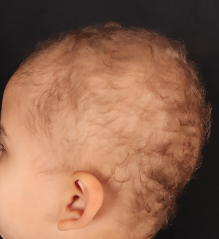
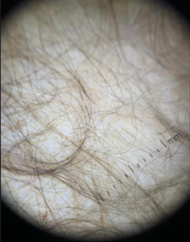
There were no abnormalities in the patient’s biochemical values. The patient had no growth retardation.
Due to complaints related to vision, she was referred to the ophthalmology department. During her examination, the best-corrected visual acuity was evaluated as 0.7 logMAR in both eyes, without relative afferent pupillary defect. The patient had no pupillary or anterior segment abnormalities, and extraocular movements were normal. Intraocular pressure was measured as 18 mmHg in the right eye and 19 mmHg in the left eye. Dilated fundus examination revealed bilateral pigmentary changes at the fovea (, ). Optical coherence tomography (OCT) of the patient showed loss of the outer retinal layers (, ).
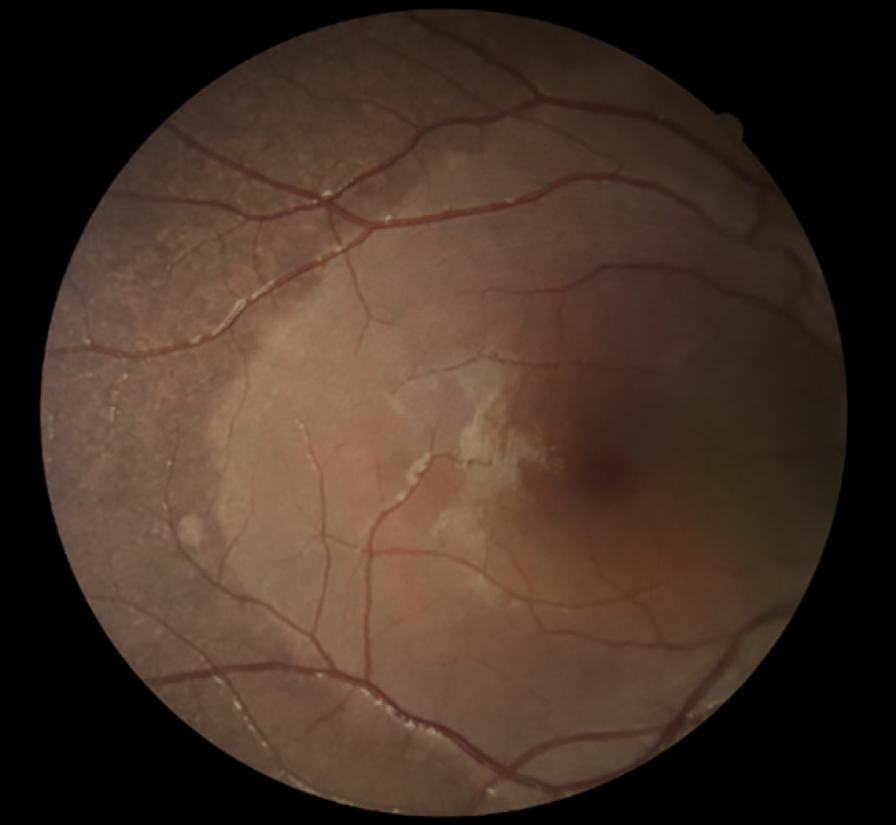
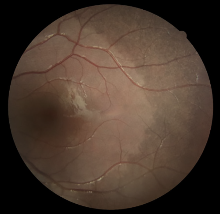
The clinical findings and complaints of the patient raised suspicion for the diagnosis of HJMD, and the patient was referred to the medical genetics department. All exons of genes and exon-intron junction regions, using library products prepared with the twist clinical exome sequencing (CES) kit from DNA samples isolated from peripheral blood, were amplified by polymerase chain reaction, followed by next-generation DNA sequencing analysis on the MGI DNBSEQ-G400 instrument.
As a result of CES, the homozygous c.830del(p.Gly277AlafsTer20) variant in the CDH3(NM_001793.6) gene was detected and this variant has been previously described in the literature. It has been reported as pathogenic in ClinVar on five occasions (RCV001851938). The variant has not been observed in population frequency studies. According to in silico assessment tools, it is damaging. Based on the available information, this variant is classified as pathogenic according to the American College of Medical Genetics and Genomics guidelines (PVS1, PM2, PM3, PP5).
Our clinical suspicion in the patient was confirmed by genetic analysis, and the patient’s family was referred for genetic counseling. The absence of other anomalies besides hypotrichosis and retinal dystrophy excluded ectodermal dysplasia, ectrodactyly, and macular dystrophy, collectively known as Ectodermal dysplasia-ectrodactyly-macular dystrophy syndrome. No clinical pathology was detected in the patient’s two siblings and mother. Nevertheless, the patient’s siblings and mother were sent for genetic analysis and the details are provided under the second case.
With written consent from the parents and verbal consent from the patient, two scalp biopsies were performed, and no specific findings were present in histopathology.
CASE 2
When the father brought his daughter for a second examination, similar hair findings were noticed in him as well.
The 45-year-old male patient also had thin and sparse hair. Additionally, he described thinning of his body hair. He also complained of vision loss and stated that he had been diagnosed with “retinitis pigmentosa”.
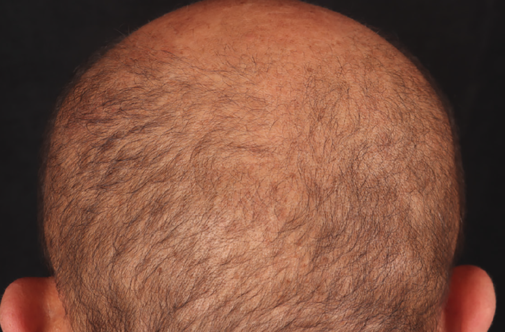
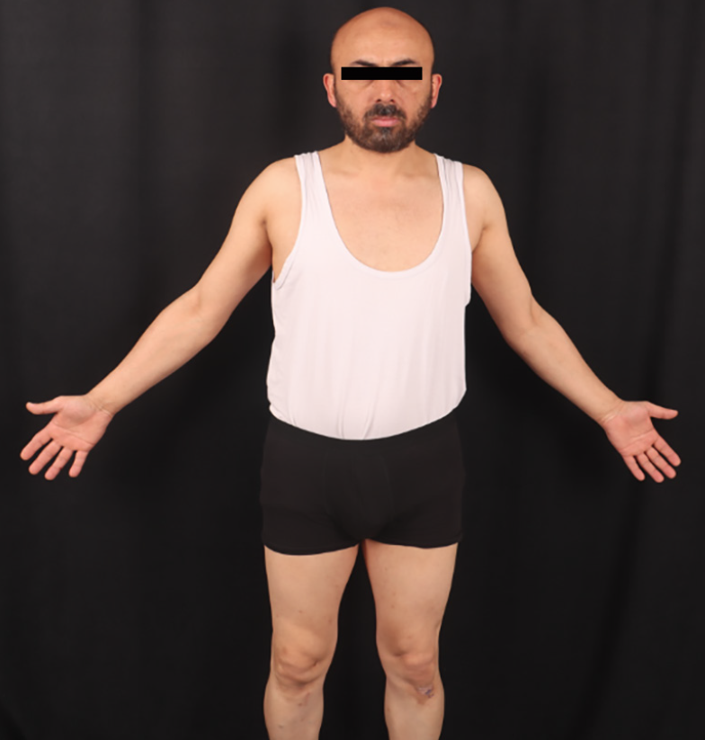
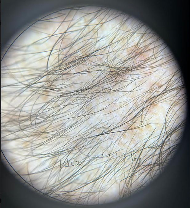
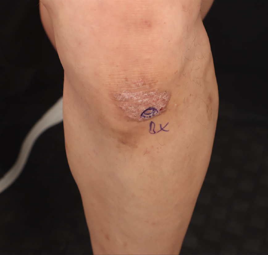
During the dermatological examination, the patient had thin, short, and sparsely distributed hair, with reduced density compared to normal (). The density of eyebrows, beard, and eyelashes was normal; however, there was decreased hair density on the upper and lower extremities, indicating hypotrichosis (). Vellus-type hair with thin, short, and low density was observed on trichoscopy over actinic damage on the scalp (). Hair pull-test was negative. On both extensor surfaces of the knees, annular erythematous and infiltrated plaques covered with silver-colored scales, with diameters of 3*3 and 4*3 cm, were observed and evaluated as psoriasis (). Examination of the teeth, nails and mucous membranes was normal.
There were no abnormalities in the patient’s laboratory values, cardiac, and other systemic examinations.
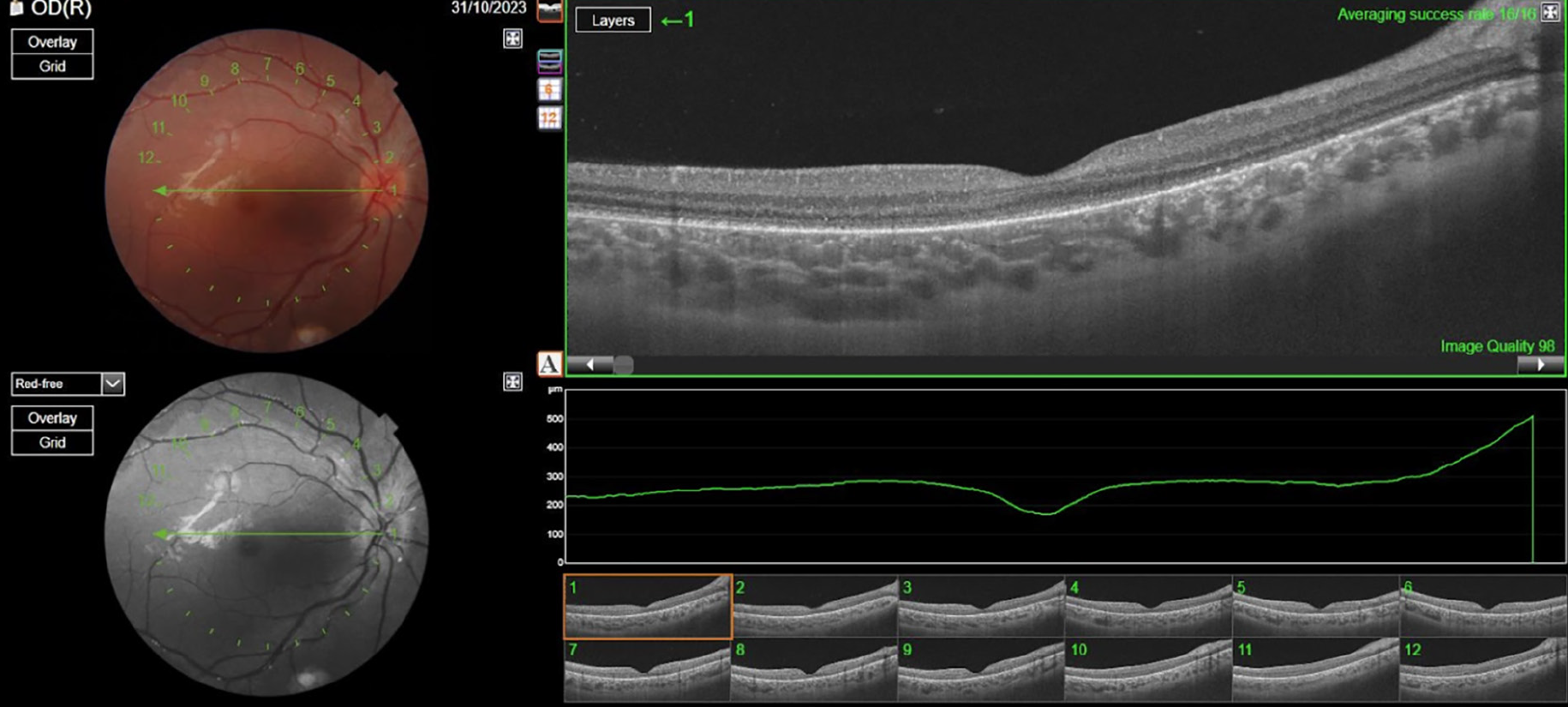
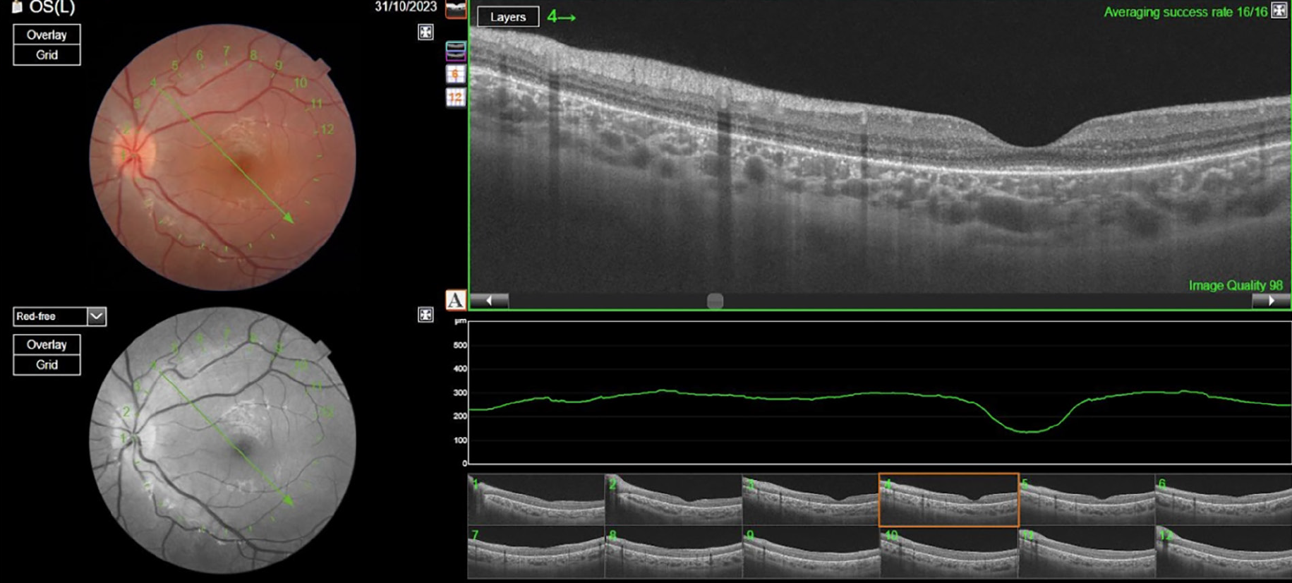
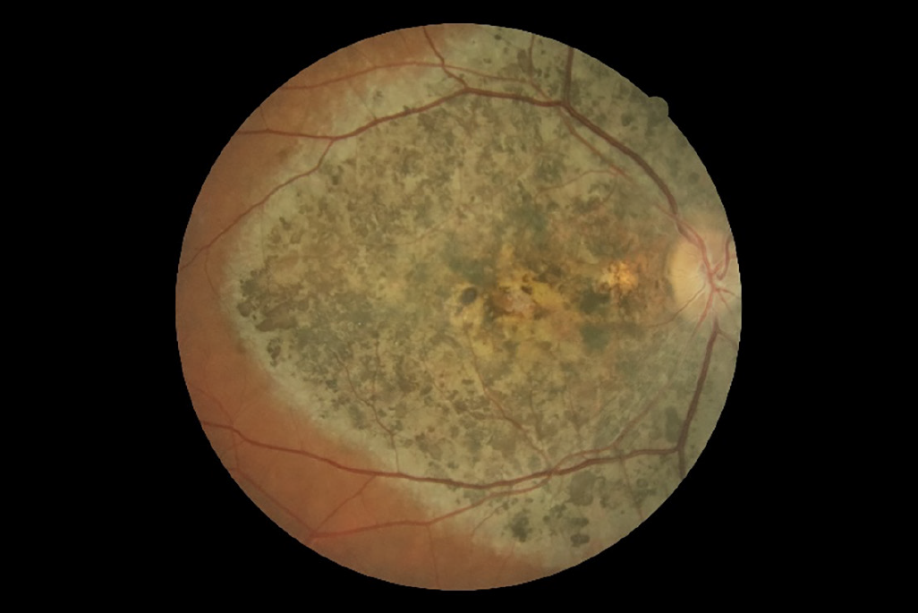
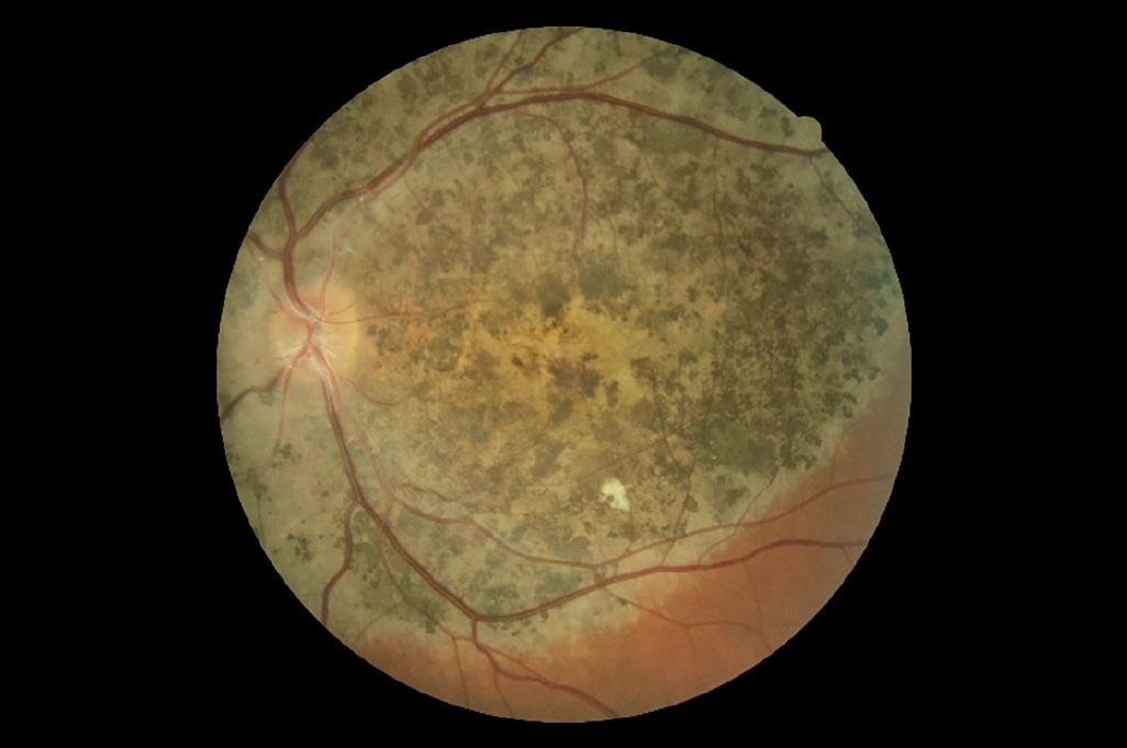
He was referred to the ophthalmology department, and his best-corrected visual acuity was 0.8 logMAR in the right eye and 1.7 logMAR in the left eye. Intraocular pressure was 14 mm Hg in the right eye and 12 mm Hg in the left eye, as measured with a Goldmann applanation tonometer. Biomicroscopic examination revealed no anterior segment pathology. Fundus examination showed marked pigmentary changes in the posterior pole (, ). In OCT, bilateral photoreceptor layer irregularity and retinal pigment epithelium atrophy, full-thickness macular hole on the right eye (), and foveal atrophy on the left eye () were detected.
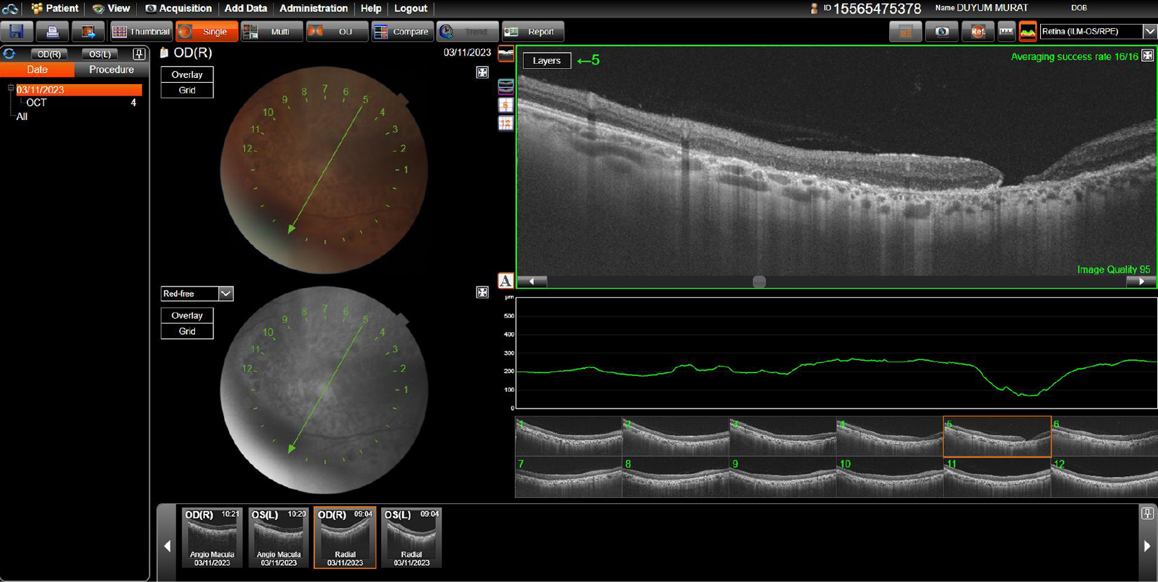
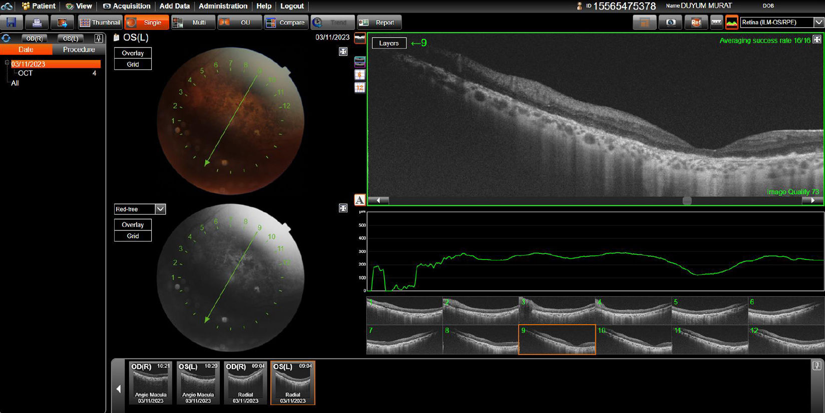
A decrease in visual acuity and bilateral irregularities in the retinal pigment epithelium were observed. Based on these findings, the patient was referred to the genetics department for genetic studies with a suspicion of HJMD. As a result of the known mutation analysis performed by Sanger sequencing, the c.830del (p.Gly277AlafsTer20) variant in the CDH3 was also detected as homozygous. Thus, the patient was also diagnosed with HJMD.
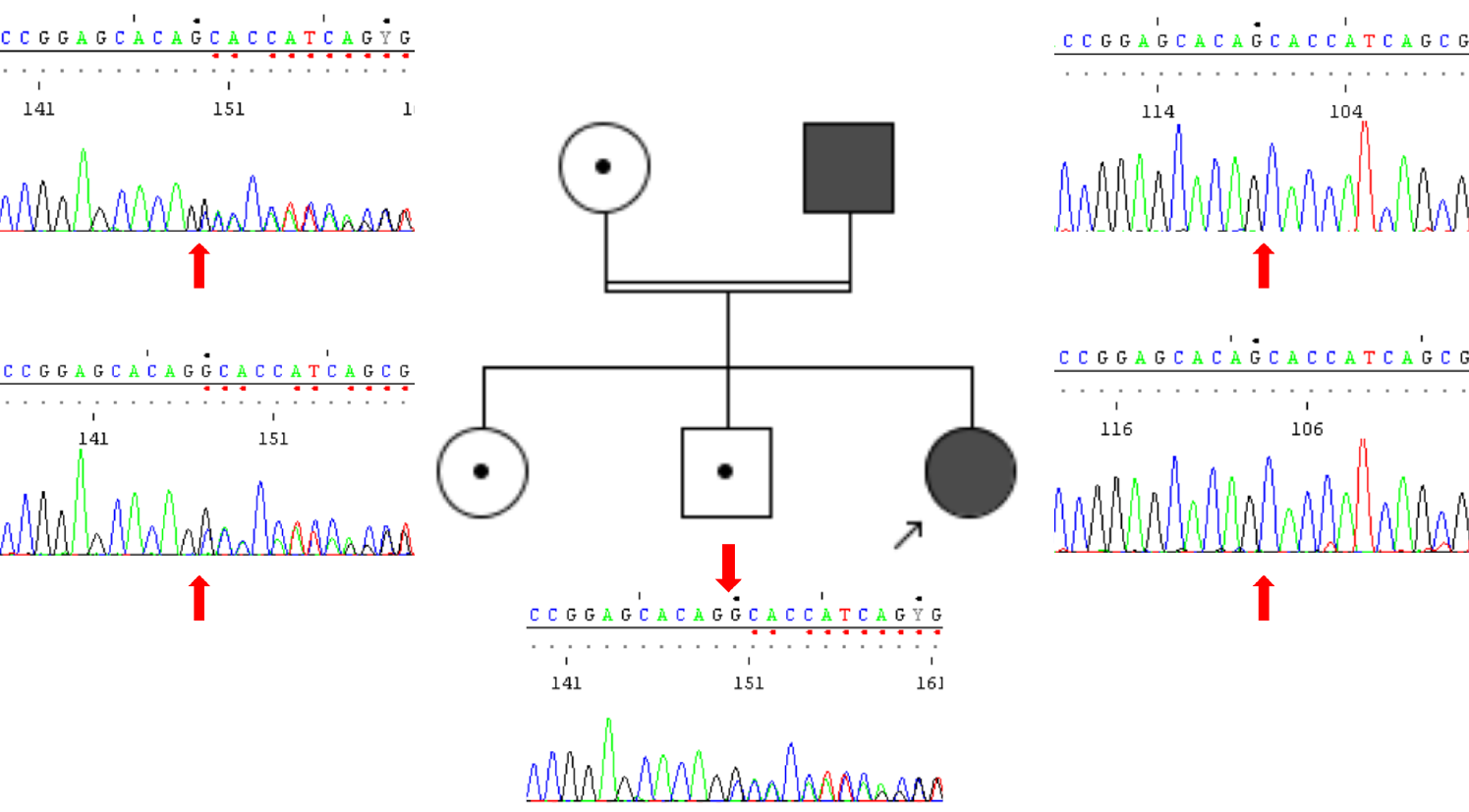
Subsequent Sanger segregation studies for the same variant of the unaffected children of the father and the mother of the index case (above-mentioned 4-year-old girl) revealed that both the unaffected children and the mother were heterozygous carriers of the variant ().
The patient consented, and biopsies from the scalp and a psoriatic plaque revealed findings consistent with psoriasis, with no specific results from scalp samples.
DISCUSSION
HJMD is a rare autosomal recessive disease first described in 1935, which can result in blindness between the ages of 20 and 40. Nearly all patients have had short, sparse hair since birth and progressive macular degeneration. The condition typically affects the hair and does not affect other body hair, however, it can sometimes affect the eyebrows and eyelashes as well. The thinning of body hair observed in our second patient suggests that this condition may affect body hair. Due to clinical suspicion, in the examination conducted on the father of our index patient, the absence of body hair emerges as a significant finding. Unlike what is known, body hair may also be affected, suggesting that this issue may need further investigation.
This disease is associated with mutations in the CDH3 gene, which encodes P-cadherin expressed in the retinal pigment epithelium and hair follicles.
The retinal component of this syndrome has been re-evaluated, revealing earlier ocular involvement than previously suggested in its previous description as “juvenile”. Contemporary investigations have demonstrated a wider retinal impact extending beyond macular involvement. In the two cases presented, fundus photography revealed various alterations in the retinal pigment epithelium, notably, accentuating the axial reflex. Both cases exhibited a circular pigmented area indicative of chorioretinal atrophy upon color fundus examination. Conversely, in case 2, OCT showed backscattering phenomena alongside diverse changes in the retinal pigment epithelium and photoreceptor layer. To the best of our knowledge, this marks the initial observation of a macular hole within the diagnosis of HJMD, shedding new light on the syndrome’s ocular manifestations.
The diagnosis of the girl, which is very rare and based on clinical suspicion, was made alongside that of her father. The father was previously misdiagnosed with “retinitis pigmentosa” due to decreased vision. To the best of our knowledge, this is the first reported father-daughter case of HJMD from Türkiye, thereby contributing novel data to the existing literature.
HJMD currently lacks a definitive treatment. In cases where hypotrichosis raises suspicion and screenings reveal ocular findings inconsistent with typical macular degeneration, thorough systemic evaluation is crucial. Such patients may exhibit sparse scalp hair alongside an underlying genetic disorder. Effective diagnosis and prognosis of conditions like HJMD require close collaboration between dermatologists, ophthalmologists, and medical geneticists. Regular follow-up is essential to monitor and manage the likelihood of the condition progressively deteriorating over time.
HJMD, a very rare disorder with approximately fifty reported cases in the literature, is characterized by hypotrichosis and progressive macular degeneration and leads to blindness within the first thirty years of life. In daily practice, differentiating diagnoses for patients with hair disorders, such as hair loss, and conducting genetic analysis when necessary are crucial for diagnosing genetic diseases associated with hair abnormalities. As in this case, although there is no definitive treatment for these diseases, providing genetic counseling for affected individuals and their families to guide future reproductive decisions as well as monitoring patients for potential effects are important. In our index case, the father, who brought the patient for examination, also exhibited hair-related findings and described having an “eye disorder,” leading to a diagnosis through genetic testing. Additionally, while HJMD typically causes hypotrichosis on the scalp, it rarely affects the eyebrows, eyelashes, or body hair, and involvement is generally not expected. However, the presence of reduced body hair in the father is a significant finding. Furthermore, the coexistence of psoriasis in the father suggests that additional clinical features may be present within the spectrum of this disease. Moreover, the identification of a macular hole in the father adds a novel ocular manifestation to the disease spectrum, broadening the current understanding of CDH3-related disorders. This paper is significant because it contributes to the literature with the first reported father-daughter HJMD case from Türkiye.
Footnotes
Ethics
Ethics Committee Approval: Ethics Committee approval was obtained from the İstanbul Kent University Ethics (approval number: E-10420511-051-34424, date: 02.07.2024).
Informed Consent: Written consent for publication of the cases including the photographs was obtained.
Authorship Contributions
Surgical and Medical Practices: N.C., Concept: N.C., Design: N.C., Data Collection or Processing: N.C., A.G., M.K., E.M.K., E.Ç., H.İ.Y., Analysis or Interpretation: N.C., A.G., M.K., Literature Search: N.C., A.G., M.K., Z.T, Writing: N.C., A.G., M.K., Z.T.
Conflict of Interest: The authors declared that they have no conflict of interest.
Financial Disclosure: The authors declared that this study received no financial support.
References
Liu Y, Xu Z, Sun Y, Zhang B, Wang X, Ma L. Hypotrichosis with juvenile macular dystrophy with novel mutations inCDH3 gene. Australas J Dermatol. 2024;65:91-94.
Almeida FT, Carneiro-Freitas R, Caldas R, Vieira AP. Hypotrichosis with Juvenile Macular Dystrophy. Int J Trichology. 2018;10:234-236.
Oliveira-Ferreira C, Leuzinger-Dias M, Tavares-Ferreira J, Silva SE, Brandão E, Falcão-Reis F, Rocha-Sousa A. Hypotrichosis with juvenile macular dystrophy. Ophthalmic Genet. 2019;40:574-577.
Richards S, Aziz N, Bale S, Bick D, Das S, Gastier-Foster J, Grody WW, Hegde M, Lyon E, Spector E, Voelkerding K, Rehm HL; ACMG Laboratory Quality Assurance Committee. Standards and guidelines for the interpretation of sequence variants: a joint consensus recommendation of the American College of Medical Genetics and Genomics and the Association for Molecular Pathology. Genet Med. 2015;17:405-424.
Ahmed A, Alali A, Alsharif O, Kaki A. Hypotrichosis with Juvenile Macular Dystrophy in Saudi Arabia: A Case Report. Skin Appendage Disord. 2021;7:75-79.
Indelman M, Eason J, Hummel M, Loza O, Suri M, Leys MJ, Bayne M, Schwartz FL, Sprecher E. Novel CDH3 mutations in hypotrichosis with juvenile macular dystrophy. Clin Exp Dermatol. 2007;32:191-196.
Rishi E, Goel S, Bassi S, Rishi P. Hypotrichosis with Juvenile Macular Dystrophy in a Patient with Cadherin 3 (CDH3) Mutation. Nepal J Ophthalmol. 2022;14:168-172.
Blanco-Kelly F, Rodrigues-Jacy da Silva L, Sanchez-Navarro I, Riveiro-Alvarez R, Lopez-Martinez MA, Corton M, Ayuso C. New CDH3 mutation in the first Spanish case of hypotrichosis with juvenile macular dystrophy, a case report. BMC Med Genet. 2017;18:1.