
Abstract
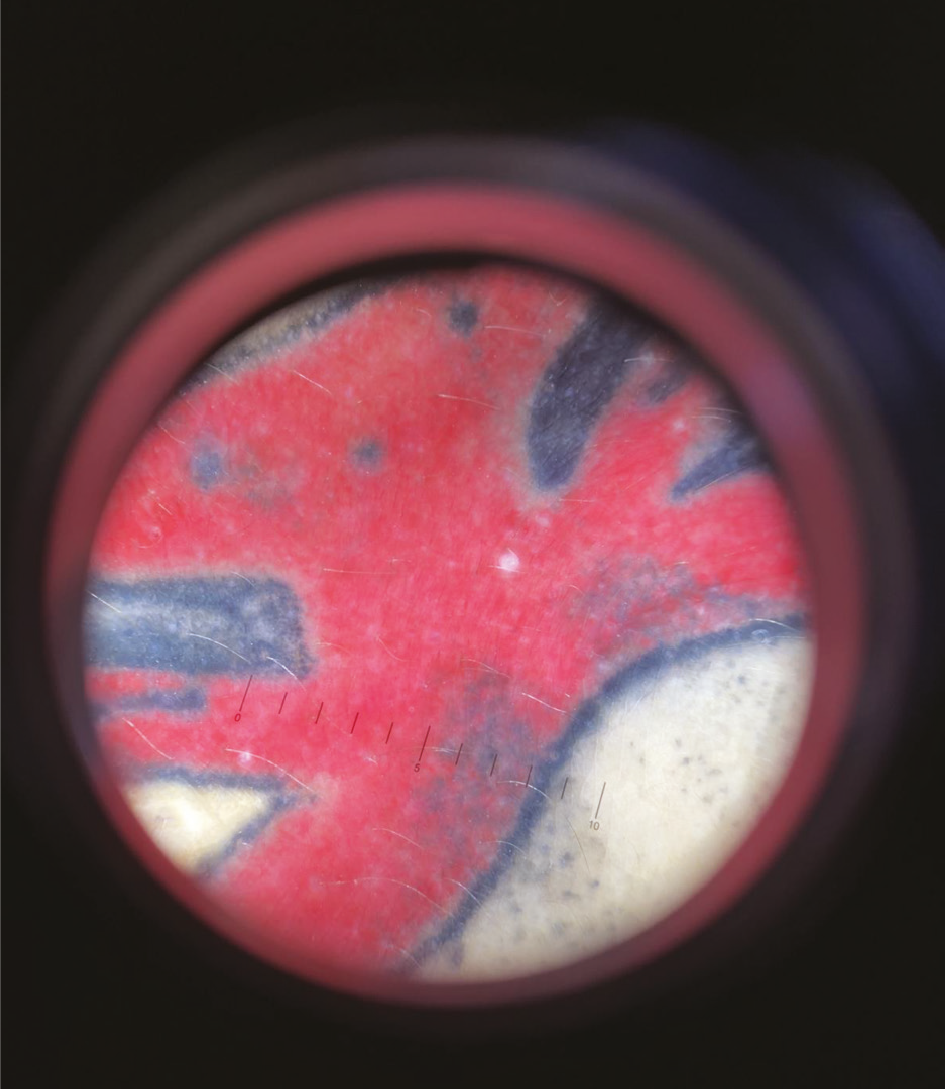
Eruptive milia within tattoos are rare and usually self limited, yet they can mimic tattoo related inflammatory reactions that require aggressive treatment. We report a 21 year old man with tiny white papules confined to the red ink portions of a recent tattoo; dermoscopy showed multiple, millimetric, bright-white round clods, without a specific vascular component; with a smooth surface and no scale over a homogeneous red background supporting a diagnosis of milia. Recognizing this benign entity and distinguishing it from red pigment reactions such as lichenoid, ranulomatous/sarcoidal, or pseudolymphomatous changes helps avoid unnecessary treatments (e.g.,corticosteroids, excision or laser therapy).
Keywords: Eruptive, milia, tattoo, red pigment
INTRODUCTION
Tattoo-related dermatoses include allergic (eczematous), granulomatous, lichenoid, photosensitive, pseudolymphomatous, and infectious reactions, collectively affecting approximately 2% of tattooed individuals.¹ Eruptive milia within tattoos are a rare complication; only a few cases have been reported, and a recent review identified eight instances of tattoo-associated milia.² Milia in tattoos are thought to result from follicular occlusion or epidermal injury caused by ink injection, possibly accentuated by hypersensitivity to pigment components., Red ink (cinnabar) is most frequently implicated in tattoo-related reactions, and lichenoid responses to red pigment have occasionally included secondary milia. Herein, we describe a rare case of eruptive milia arising in a red-ink tattoo, highlighting its dermoscopic features and summarizing previously reported cases to help clinicians in distinguishing this benign entity from red pigment-related inflammatory reactions.
CASE REPORT
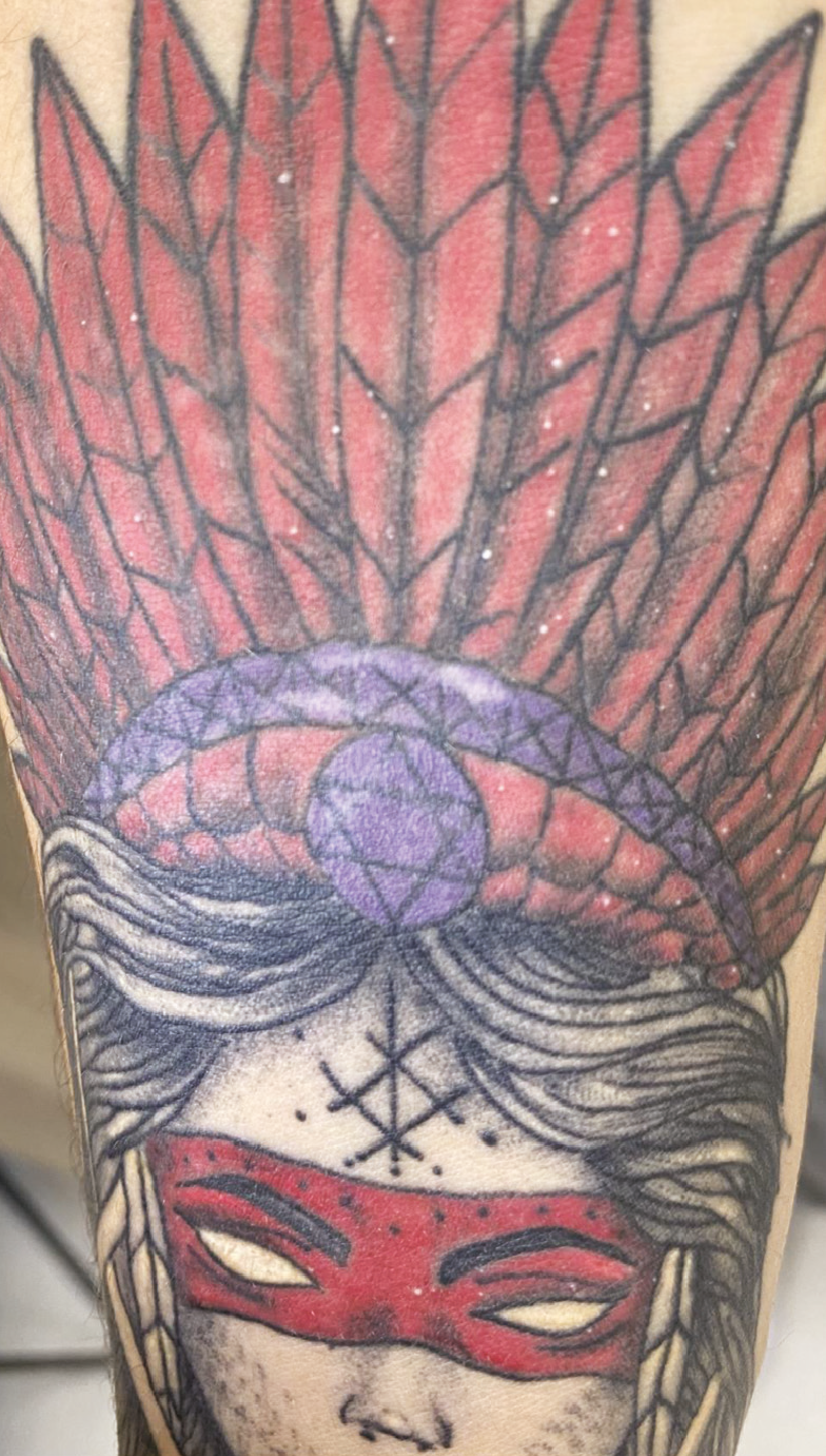
A healthy 21-year-old man presented with asymptomatic white papules confined to the red-ink portions of a newly applied forearm tattoo; the papules appeared one month after its completion. The patient used only dexpanthenol cream for aftercare. Clinical examination of his right forearm revealed multiple millimetric, white, dome-shaped papules scattered across the red-pigmented areas, sparing the surrounding skin (). Dermoscopy showed multiple, millimetric, bright-white, round clods with a smooth surface and no scale, set on a homogeneous red background and lacking a specific vascular component (). Based on these clinical and dermoscopic findings, a diagnosis of eruptive milia within the tattoo was made. Topical therapy with 0.025% tretinoin cream was initiated nightly. The patient was scheduled for a three-month follow-up; however, he was lost to follow-up, and the treatment outcome could not be assessed.
DISCUSSION
Eruptive milia that develop within tattoos are uncommon, yet increasingly recognized in the dermatologic literature., Lesions typically emerge within weeks to a few months after tattooing and may occur with various pigments, though red ink is most often implicated. Red pigments, frequently containing metals or azo dyes (e.g., mercury, cadmium, ferric hydrate), are well known to provoke immunologic reactions such as lichenoid, granulomatous, or pseudolymphomatous processes, whereas black carbon-based inks are largely inert.
Only eight cases of tattoo-related eruptive milia have been reported to date, commonly involving red or other bright pigments such as yellow or green., Details of these cases, including patient demographics, pigment color, onset time, treatment, and outcome are summarized in ., , - Reported lesions were either limited to specific color areas or distributed throughout multicolored tattoos while sparing adjacent normal skin.
Table 1. Characteristics of published cases of eruptive milia associated with tattoos
Author (year) | Age/sex | Pigment color | Onset time after tattoo | Treatment | Outcome/notes |
|---|---|---|---|---|---|
Lucke et al., | 36, male | Red (milia associated with lichenoid reaction) | - | N/A | N/A |
Koh et al., | 24, male | Multiple colors | 1 month | Urea 10% + salicylic acid 2% | Mild improvement |
Miller et al., | 28, male | Red and yellow | 3 months | None | Near‑complete spontaneous resolution at 6 months |
Ross et al., | 19, female | Multiple colors | 1 month | Urea 40% + tretinoin 0.1% | Resolved by 2 months |
Uraga et al., | 26, female | Red, blue | 5 months | Not stated | Not stated |
Kluger, | 32, male | Red, black, green | 2.5 months | Topical tretinoin | Outcome not reported |
Kluger, | N/A, female | Grey | N/A | N/A | N/A |
Kluger, | N/A, male | Black | N/A | N/A | N/A |
Present case | 21, male | Red | 1 month | Topical tretinoin | Patient lost to follow-up |
N/A: Not available
The pathogenesis remains incompletely understood but appears multifactorial, primarily related to mechanical trauma and adnexal injury. Repetitive needle penetration induces an acute, aseptic inflammatory reaction that can damage the follicular infundibulum or eccrine ducts, predisposing to secondary cyst formation., Histopathologic findings from a previously reported lichenoid tattoo reaction with eruptive milia demonstrated partial destruction of adnexal structures, supporting this proposed mechanism. Post-tattoo care measures, such as occlusive ointment massage, may promote keratin retention within micro-wounds and follicular ostia, contributing to milia formation. Other secondary causes of milia, including burns, dermabrasion, and ablative laser procedures, reinforce the role of epidermal trauma and abnormal keratinization. Although hypersensitivity to pigment components, especially red dyes, may induce localized inflammation, current evidence favors trauma-induced adnexal disruption and keratin accumulation as the main pathogenic mechanism.
While the diagnosis of milia is generally clinical, differentiation from other post-tattoo papular eruptions, such as granulomatous, pseudoepitheliomatous, pseudolymphomatous, or lichenoid reactions, is crucial. Dermoscopy showing tiny bright-white round clods on a smooth, non-scaly surface without a vascular component supports the diagnosis and helps avoid unnecessary biopsies. Nevertheless, the dermoscopic patterns of tattoo-associated reactions have been described only in a few case reports, and systematic data remain limited. Granulomatous reactions typically display brownish-gray structureless areas with white scale, crystalline structures, and branching or irregular vessels. Pseudoepitheliomatous hyperplasia exhibits two distinct dermoscopic zones: a central keratotic area with white scale, pink-white structureless regions, comedo-like openings, white circles, red globules, hemorrhage, and hairpin or irregular vessels, surrounded by a peripheral gray-to-bluish-white zone.
Tattoo-associated pseudolymphoma demonstrates a homogeneous violaceous to violet-pink background with ill-defined borders and occasional white-yellow perifollicular halos. In lichenoid tattoo reactions, dermoscopy reveals diffuse white scaling, pink-white structureless areas, comedo-like openings keratotic plugs, shiny white lines, rosettes, and linear or irregular vessels, but not the blue-white or gray-white areas and Wickham’s striae seen in idiopathic lichen planus.
Although biopsy was not performed in our patient, previously reported cases describe small keratin-filled cysts lined by stratified squamous epithelium, containing concentrically laminated keratin. Tattoo pigment granules are commonly seen extracellularly in the dermis and within dermal macrophages, while spongiosis and inflammation are absent, confirming a benign, non-inflammatory process.
Eruptive milia are benign, primarily of cosmetic concern, and may resolve spontaneously without treatment. In published cases, topical keratolytics (urea) softened the stratum corneum and released keratin plugs, whereas topical retinoids enhanced epidermal turnover and prevented new cyst formation.³ The treatments used, and their outcomes are summarized in ., , - These conservative approaches are typically curative, and manual extraction or laser therapy is reserved for resistant cases. In our patient, topical tretinoin was prescribed; however, he was lost to follow-up, and treatment response could not be documented.
CONCLUSION
Eruptive milia within tattoos are rare, benign, and self-limited lesions that may mimic inflammatory or granulomatous tattoo reactions. Recognition of their clinical and dermoscopic features prevents misdiagnosis and unnecessary interventions. Awareness of this entity, particularly in red-ink tattoos, helps clinicians distinguish it from immune-mediated tattoo complications. Although follow-up was unavailable in our case, prior reports indicate good response to topical keratolytics or retinoids, underlining the effectiveness of conservative management.
Footnotes
Ethics
Informed Consent: Written informed consent was obtained from the patient for publication of clinical details and images.
Authorship Contributions
Surgical and Medical Practices: B.Ç.C., F.K., Concept: F.H., S.P.K., Design: F.H., S.P.K., Data Collection or Processing: B.Ç.C., F.K., Analysis or Interpretation: F.H., B.Ç.C., F.K., S.P.K., Literature Search: F.H., Writing: F.H., B.Ç.C., F.K., S.P.K.
Conflict of Interest: The authors declared that they have no conflict of interest.
Financial Disclosure: The authors declared that this study received no financial support.
References
Kazandjieva J, Tsankov N. Tattoos: dermatological complications. Clin Dermatol. 2007;25(4):375-382.
Kluger N. Eruptive milia and acneiform hyperkeratosis with comedones (pseudo-epidermal cysts) within tattoos. Ann Dermatol Venereol. 2019;146(12):801-806.
Ross N, Farber M, Sahu J. Eruptive milia within a tattoo: a case report and review of the literature. J Drugs Dermatol. 2017;16(6):621-624.
Mortimer NJ, Chave TA, Johnston GA. Red tattoo reactions. Clin Exp Dermatol. 2003;28(5):508-510.
Temiz SA, Özlü E. Medical complications of tattoos. J Turk Acad Dermatol. 2021;15(1):1-7.
Lucke T, Fallowfield M, Burden D. Lichen planus associated with milia. Clin Exp Dermatol. 1999;24(4):266-269.
Koh MJ, Teo RY, Liu TT. Multiple epidermal cysts occurring in a tattoo. Singapore Med J. 2009;50(11):e376-377.
Miller LM, Schwartz JT, Cho S. Milia: a unique reaction to tattoos. Cutis. 2011;87(4):195-196.
Uraga E, Loayza E, Briones MC, Uraga V. Utilidad de la dermatoscopia en el diagnóstico de milia desarrollada sobre tatuaje previo. PIEL Latinoam. 2009 [Internet]. Available from: https://piel-l.org/blog/wp-content/uploads/2009/01/utilidad-de-la-dermatoscopia.pdf
Kalantari Y, Peymanfar AA, Mahmoudi H, Daneshpazhooh M, Etesami I. Dermoscopy of cutaneous granulomatous disorders: a study of 107 cases. Skin Res Technol. 2023;29(1):e13273.
Behera B, Kumari R, Gochhait D, Ayyanar P. Dermoscopy of pseudoepitheliomatous hyperplasia tattoo reaction pattern. Dermatol Pract Concept. 2022;12(3):e2022121.
Kendel M, Toncic RJ, Bradamante M, Ilic I, Loncaric D, Rados J, Drvar DL. Dermoscopy of a tattoo pseudolymphoma. Dermatol Pract Concept. 2019;9(1):17-19.
Sethy M, Behera B, Dash S, Palit A, Nayak AK, Ayyanar P. Clinicodermoscopic and immunopathological profile of non-infectious non-eczematous inflammatory tattoo reactions: a retrospective study from a tertiary care centre of East India. Indian J Dermatol Venereol Leprol. 2023;89(4):558-567.