
Abstract
Aim
Mycosis fungoides [(MF)— the most common primary cutaneous T-cell lymphoma —] is difficult to diagnose early, and treatment access is uneven in middle-income settings; therefore, we aimed to describe current practice, identify barriers, and propose guideline-aligned, resource-adapted solutions.
Materials and Methods
Cross sectional anonymous online survey of dermatologists (May–June 2025) on diagnostic workflows/access, treatments, and barriers. Analyses were performed using SPSS v23, with descriptive statistics and Spearman’s rho correlations. Two-tailed P < 0.05 was considered significant.
Results
Among 239 respondents, 61.1% managed MF; 49.3% reported diagnostic uncertainty, and T-cell receptor testing was rarely available. The use of diagnostic algorithms and structured training was inconsistent. Basic topical agents and retinoids were widely available, whereas advanced systemic and device-based options were scarce. Barriers clustered around registration and market availability, workforce constraints, and equipment and maintenance issues. Reported workarounds included evidence-based substitutions, interim symptom-directed therapy, repeat biopsies, and referrals; multidisciplinary tumor boards were underused.
Conclusion
MF care is heterogeneous and resource constrained. A four-component plan — a national diagnostic algorithm with a minimum package and a re-biopsy–consultation–multidisciplinary team loop; targeted capacity building; tiered treatment pathways prioritizing narrow-band ultraviolet B ± retinoids with clear referral thresholds; and system integration via centers of excellence, a national registry, and device uptime programs — may standardize care and improve outcomes.
Keywords: Mycosis fungoides, cutaneous T-cell lymphoma, diagnostic challenges, therapeutic accessibility, resource-adapted solutions
INTRODUCTION
Mycosis fungoides (MF) is the most common form of primary cutaneous T-cell lymphoma (CTCL), representing the majority of CTCL cases worldwide.¹ It typically presents as slowly evolving erythematous or mildly pigmented, atrophic patches in sun-protected sites and may progress to infiltrated plaques, tumors, or erythroderma in advanced stages., MF displays substantial clinicopathologic heterogeneity, making early diagnosis difficult. Lesions may mimic other dermatoses, such as eczema, psoriasis, or chronic dermatitis, and histopathologic findings are often non-specific., In this context, clinicopathological correlation, supported by immunophenotyping and, where available, molecular testing, is essential, and multiple international guidelines recommend a multimodal diagnostic approach. However, real-world adherence to these recommendations varies widely depending on access to diagnostic resources, specialist expertise, and healthcare infrastructure.
Since accurate diagnosis forms the foundation for disease staging and staging is the principal determinant of treatment selection, these processes are intrinsically linked to ensuring optimal MF care. Early-stage disease is generally approached with skin-directed therapies, while advanced stages often require systemic agents, frequently in combination with skin-directed therapies for symptom control.- The therapeutic hierarchy outlined in international guidelines is supported by systematic reviews and meta-analyses, which confirm the efficacy of these modalities but also reveal substantial variation in patient outcomes and treatment availability across healthcare systems.- Such differences are often shaped by disparities in access to advanced diagnostic tools, limitations in the availability of systemic therapies, and inconsistent application of guideline-based protocols, all of which may lead to delays in diagnosis and deviations from evidence-based management.
We hypothesized that significant variability exists in both diagnostic and therapeutic practices for MF, shaped by disparities in resource availability and guideline implementation. To investigate this, we conducted a nationwide, cross-sectional survey among dermatologists in a middle-income country with universal health coverage, aiming to identify current practice patterns, barriers to optimal care, and locally adapted solutions to improve MF management within existing resource constraints.
MATERIALS AND METHODS
Survey Design
This cross-sectional, questionnaire-based study was conducted between May and June 2025 to evaluate current challenges and potential solutions regarding diagnosis and access to treatment for MF among dermatologists in Türkiye. The survey instrument was developed by the study team. It consisted of qualitative and quantitative items, including open-ended, multiple-choice, and multiple-selection questions. The questionnaire aimed to investigate dermatologists’ clinical experience with MF, diagnostic approaches, access to diagnostic modalities (e.g., histopathology, immunohistochemistry, molecular testing), treatment preferences (topical or device-based skin-directed therapies, systemic therapies), and barriers encountered in treatment access (e.g., reimbursement, availability, institutional limitations). Institutions were classified by service scope and technical capacity; rankings were based on technical resources, patient volume, and academic staff, in the following order: University Hospital, Training and Research Hospital, City Hospital, State Hospital, and Private Clinic. The content of the questionnaire is presented in . The final version of the questionnaire was created using Google Forms. A survey link was generated and disseminated to Turkish dermatologists through dermatology-focused professional WhatsApp and Facebook groups on three separate occasions at two-week intervals, to enhance response rates and ensure representative participation.
Participants
The target population comprised dermatologists actively practicing in Türkiye who manage patients with MF in various clinical settings. Informed consent was obtained digitally prior to survey participation. The first page of the online survey included an information section outlining the purpose of the study, data use, and confidentiality. Only participants who provided digital consent could proceed to complete the remaining parts of the questionnaire. Respondents not managing MF patients were included only in descriptive analyses; items requiring direct MF management experience were analyzed within the subgroup actively managing MF.
Statistical Analysis
All responses were collected anonymously. Data were exported to IBM SPSS Statistics version 23 for analysis. Descriptive statistics were used to summarize the data, with categorical variables presented as frequencies and percentages. Correlations between ordinal variables were assessed using Spearman’s rank correlation coefficient (rho). All statistical tests were two-tailed, and a P-value < 0.05 was considered statistically significant.
RESULTS
Participant Characteristics
A total of 239 dermatologists participated in the study. shows the distribution of participants by gender, professional experience, institution type, and academic title. Among them, 38.9% (n = 93) did not manage MF patients, most often due to inadequate technical resources (57.0%, n = 53), a preference to refer patients to specialized centers (43.0%, n = 40), no MF patient admissions (32.3%, n = 30), limited clinical experience (10.8%, n = 10), or lack of interest in MF care (7.5%, n = 7).
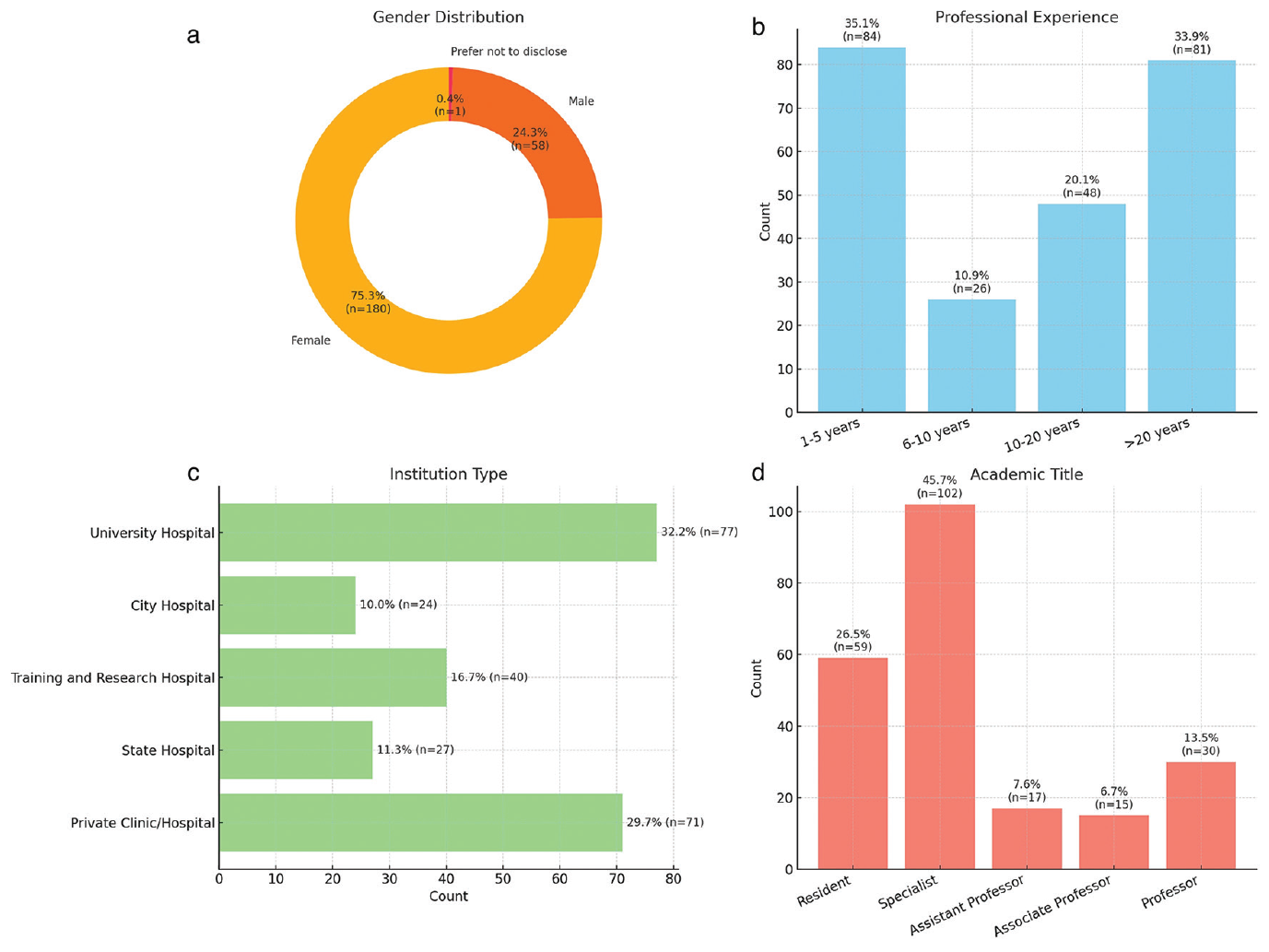
The remaining 61.1% (n = 146) reported actively managing MF patients. Most followed 0–5 patients annually (35.6%, n = 52), followed by 6–10 patients (22.6%, n = 33), 11–20 patients (13.7%, n = 20), and > 20 patients (28.1%, n = 41). Patient volume correlated positively with years of clinical experience (Spearman’s ρ = 0.45, P < 0.001). In daily practice, participants primarily referenced European Organisation for Research and Treatment of Cancer (EORTC) guidelines (35.6%, n = 52), National Comprehensive Cancer Network (NCCN) guidelines (30.8%, n = 45), or national experience (21.2%, n = 31), with relatively similar proportions across these three sources.
Diagnostic Approaches, Challenges, and Solutions
summarizes the reported diagnostic modalities. Clinical examination, multiple or repeat biopsies, and basic immunohistochemistry were used by more than 85% of respondents, whereas advanced tests, such as human T-cell lymphotropic virus-1/2 serology and T-cell receptor (TCR) gene rearrangement, were rarely or never applied.
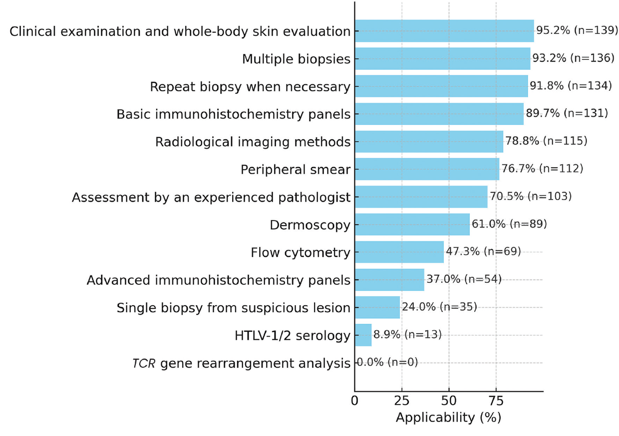
MF: Mycosis fungoides, TCR: T-cell receptor, HTLV-1: Human T-lymphotropic virus type 1, HTLV-2: Human T-lymphotropic virus type 2
Diagnostic challenges were common: 49.3% (n = 72) reported diagnostic uncertainty, and 41.1% (n = 60) did not use diagnostic algorithms or were unfamiliar with them. The most frequent obstacles were non-specific histopathology (79.5%, n = 116), atypical presentations (54.8%, n = 80), and clinicopathological discrepancies (51.4%, n = 75). Other reported barriers included pathologist inexperience (48.6%, n = 71) and limited access to advanced molecular techniques (19.2%, n = 28).
When facing such challenges, dermatologists most often performed clinical re-evaluation at follow-up (72.6%, n = 106) or performed repeated biopsies (69.9%, n = 102). Other strategies included symptomatic or topical treatment (49.3%, n = 72), pathology consultation (37.7%, n = 55), multidisciplinary discussion (19.2%, n = 28), and referral to specialized centers (24.7%, n = 36). The specific links between diagnostic barriers and adopted solutions are illustrated in .
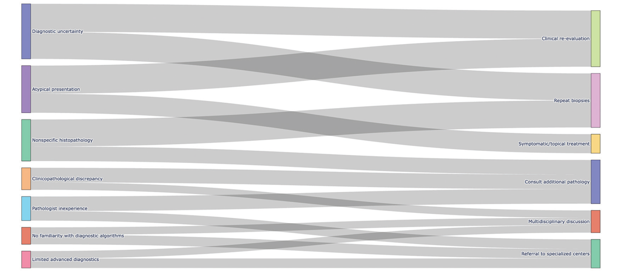
(For clarity, major diagnostic challenges and corresponding management strategies illustrated in the figure are also summarized in the main text). MF: Mycosis fungoides
Therapeutic Accessibility Challenges and Solutions
Treatment accessibility varied by category, as demonstrated in . Among topical agents, corticosteroids (99.3%, n = 145), tacrolimus (89.7%, n = 131), pimecrolimus (88.4%, n = 129), and topical bexarotene (82.2%, n = 120) were widely available, while mechlorethamine (2.1%, n = 3) and carmustine (0.7%, n = 1) were rarely or never accessible.
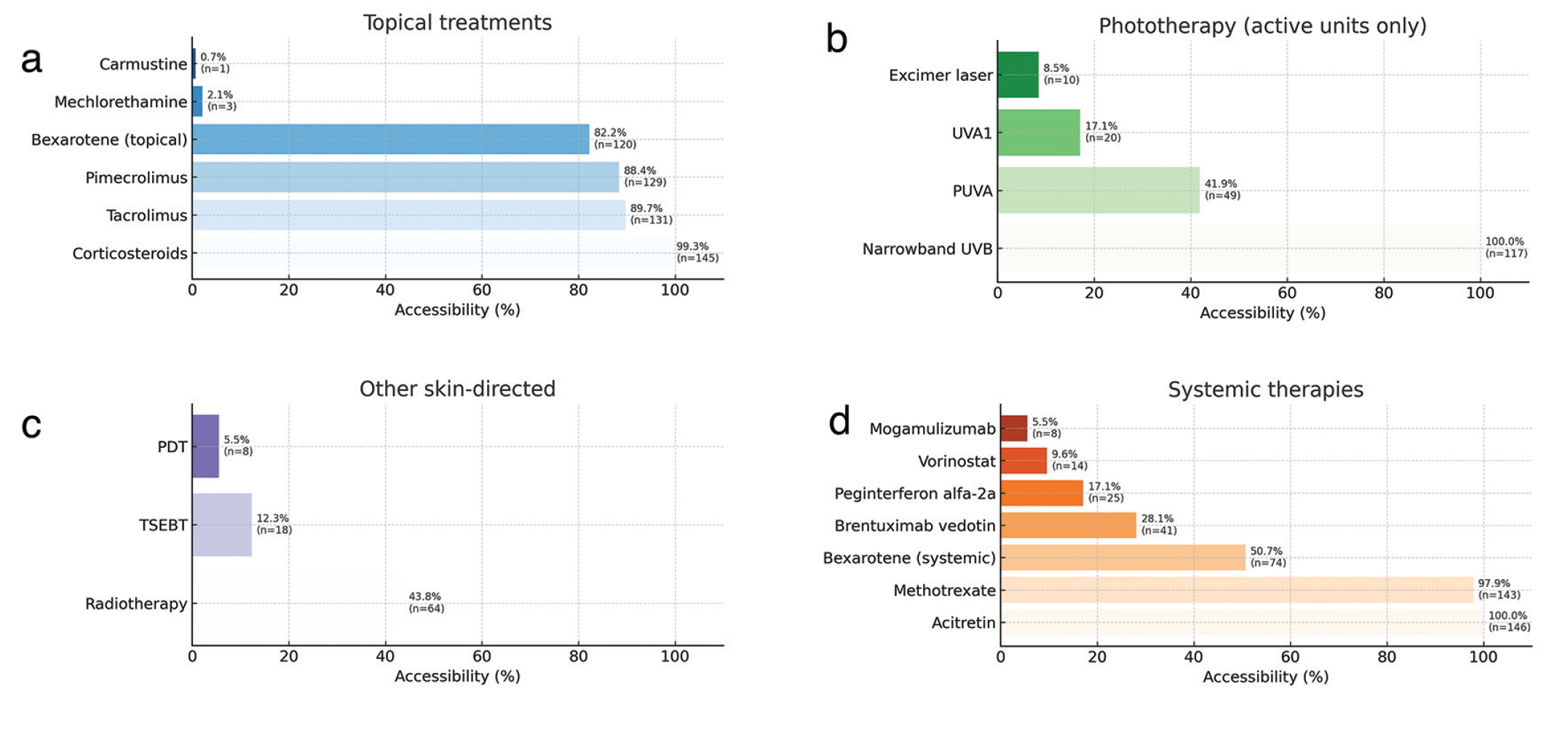
MF: Mycosis fungoides, PUVA: Psoralen ultraviolet A, TSEBT: Total skin electron beam therapy, PDT: Photodynamic therapy, UVB: Ultraviyole B
Among participants, 78.5% (n = 117) had an active phototherapy unit; narrowband ultraviolet B was universally available in these centers (100.0%, n = 117), whereas psoralen ultraviolet A (PUVA) (41.9%, n = 49), ultraviolet A1 (17.1%, n = 20), and excimer laser (8.5%, n = 10) were less common. Radiotherapy was accessible to 43.8% (n = 64) of respondents, whereas total skin electron beam therapy (TSEBT) (12.3%, n = 18) and photodynamic therapy (PDT) (5.5%, n = 8) were accessible to only a minority.
Among systemic therapies, acitretin (100.0%, n = 146) and methotrexate (97.9%, n = 143) were almost universally accessible; bexarotene was moderately accessible (50.7%, n = 74); and advanced agents such as brentuximab vedotin (28.1%, n = 41), pegylated interferon alfa-2a (17.1%, n = 25), vorinostat (9.6%, n = 14), and mogamulizumab (5.5%, n = 8) were rarely or never accessible.
Reported barriers to access included lack of product registration (80.0%, n = 120), market unavailability (75.0%, n = 112), and limited clinical experience (60.0%, n = 90) for topical agents; lack of personnel (70.0%, n = 100) or device malfunction (65.0%, n = 93) for phototherapy; and lack of knowledge/experience (55.0%, n = 80) or market unavailability (50.0%, n = 73) for systemic treatments.
Accessibility limitations often led dermatologists to use alternative therapies (70.5%, n = 103) or to refer patients to better-equipped centers (57.5%, n = 84), with referral decisions guided by proximity to those centers, academic expertise, and technical capacity. Less common strategies included off-label or non-standardized treatments (6.8%, n = 10), observational management (4.8%, n = 7), and enrollment in clinical research (4.1%, n = 6). summarizes selected treatment pathway modifications compared with major international guidelines, indicating whether these reflect guideline-endorsed alternatives, shifts in emphasis, or adaptations primarily driven by local clinical practice.
Table 1. Summary of treatment pathway modifications in relation to major international guidelines
- | Intervention | Guideline position (EORTC 2023/NCCN 2025/BAD 2018) | Alignment classification |
|---|---|---|---|
Skin-directed topical treatments | Topical tazarotene instead of topical bexarotene | EORTC 2023: Topical tazarotene is not recommended due to limited evidence and lack of availability; topical bexarotene is not approved in Europe. NCCN 2025: Recommends topical bexarotene; tazarotene is not listed. BAD 2018: Topical bexarotene mentioned; tazarotene absent. | Non-guideline substitution (experience-based) |
Skin-directed device based treatments | Prioritizing nbUVB over PUVA in early stage | EORTC 2023: PUVA and nbUVB both recommended at same evidence level. NCCN 2025: Lists PUVA and nbUVB without priority. BAD 2018: PUVA preferred in most cases. | Preference shift within guideline options |
Systemic retinoid + nbUVB instead of PUVA | EORTC 2023: PUVA and nbUVB are both recommended at the same level; systemic retinoids can be combined with phototherapy if needed. NCCN 2025: Allows retinoid + phototherapy combinations; no strict preference over PUVA. BAD 2018: PUVA preferred; combination therapy possible but not standard first choice. | Preference shift within guideline options | |
Local RT ± systemic treatment for few tumors; TSEBT ± systemic treatment for many tumors | EORTC 2023: Matches this stage-based selection. NCCN 2025: Fully aligned with RT for limited lesions and TSEBT/systemic for widespread. BAD 2018: Similar recommendations. | Full alignment | |
For device-based therapy with no alternative: refer to higher-level center | Local practice policy | ||
Systemic therapies | Replacing oral bexarotene with oral acitretin/isotretinoin | EORTC 2023: Retinoids (acitretin, isotretinoin, bexarotene) all listed; no superiority proven. NCCN 2025: Notes acitretin and isotretinoin as alternatives to bexarotene. BAD 2018: Includes retinoids as systemic options; no strict preference. | Guideline-approved alternative |
Early use of systemic retinoids ± Peg-IFN-α in plaque/folliculotropic disease | EORTC 2023: Retinoids and Peg-IFN-α are second-line/combination; not mandated early. NCCN 2025: Lists both as systemic options, usually after failure of skin-directed therapy. BAD 2018: Similar sequencing; not first-line in most cases. | Earlier use than standard | |
IFN-β instead of IFN-α | EORTC 2023: Peg-IFN-α preferred; IFN- β not listed as option. NCCN 2025: Peg-IFN-α included; no IFN-β mention. BAD 2018: IFN-α recommended; IFN- β absent. | Non-guideline substitution (experience-based) | |
Calling vorinostat early if “no response” | EORTC 2023: HDAC inhibitors (vorinostat, romidepsin) not first-line; for refractory/advanced disease. NCCN 2025: HDAC inhibitors listed after multiple prior lines. BAD 2018: Similar to EORTC/NCCN; later-line agents. | Earlier use than standard | |
Listing BV/mogamulizumab | EORTC 2023: Strong evidence for both agents in advanced or pretreated disease. NCCN 2025: Recommends BV and mogamulizumab with supporting trial data. BAD 2018: Predates widespread approval; less emphasis. | Guideline-supported (recent agents) | |
For systemic therapy with no alternative: refer to higher-level center | - | Local practice policy |
EORTC: European Organisation for Research and Treatment of Cancer, NCCN: National Comprehensive Cancer Network, BAD: British Association of Dermatologists, PUVA: Psoralen ultraviolet A, nbUVB: Narrowband ultraviolet B, RT: Radiotherapy, TSEBT: Total skin electron beam therapy, IFN-α: Interferon alpha, Peg-IFN-α: Pegylated interferon alpha, IFN-β: Interferon beta, HDAC: Histone deacetylase, BV: Brentuximab vedotin
DISCUSSION
In this study, we evaluated differences in the implementation of diagnostic and therapeutic practices for MF among dermatologists and across centers. The results of this nationwide survey revealed substantial variability in MF diagnosis and treatment among dermatologists, with significant gaps in access to advanced diagnostic tools and therapies, frequent use of basic modalities, and adaptations in clinical practice driven by resource limitations, variable local expertise, and the absence of national guidelines.
Almost half of the respondents reported diagnostic uncertainty in MF, primarily due to non-specific histopathology and clinicopathological discrepancies. In early MF lesions, the alignment of atypical lymphocytes along the basal layer of the epidermis, the presence of numerous neoplastic lymphocytes in the epidermis, accompanied by minimal spongiosis, and papillary dermal fibrosis with characteristic wire bundle–like collagen fibers may be absent. Atypical clinical presentations were another important cause of diagnostic difficulties for dermatologists diagnosing MF. The difficulty in considering MF among preliminary clinical diagnoses stems from its ability to mimic a broad spectrum of dermatoses, ranging from common inflammatory conditions such as atopic dermatitis and psoriasis to infectious and granulomatous diseases. Similar diagnostic pitfalls have been described in multiple studies in which both dermatologists and dermatopathologists have faced challenges in differentiating early MF from benign inflammatory dermatoses in the absence of sufficient experience and repeated clinicopathological correlation.
The survey results showed that 40% of dermatologists reported being unfamiliar with diagnostic algorithms. This finding highlights the need for wider implementation of algorithm-based approaches and points to a gap in training. In 2005, the International Society for Cutaneous Lymphomas proposed a diagnostic algorithm to improve the early diagnosis of MF. This algorithm includes four criteria: clinical findings, histopathological features, immunohistochemical results, and TCR gene rearrangement. Despite its high sensitivity, the algorithm’s utility in clinical practice may be limited by its relatively low specificity. The relatively high proportion of participants unfamiliar with diagnostic algorithms underscores a gap in structured diagnostic training, echoing the literature’s call for wider implementation of standardized workflows and algorithm-based approaches.,
Limited access to advanced diagnostic tools, including TCR testing and comprehensive immunohistochemistry panels, represents a major barrier to the implementation of diagnostic algorithms in routine clinical practice, particularly in middle-income healthcare systems. In our study, the absence of TCR gene rearrangement testing likely reflects restricted access to specialized molecular laboratories and reimbursement constraints rather than indicating a lack of clinical relevance. This limitation may contribute to persistent diagnostic uncertainty, especially in early-stage disease, and hinder the full application of multimodal diagnostic approaches in real-world settings.
Evidence suggests that targeted investment in diagnostic infrastructure and specialist training can significantly enhance early-stage diagnostic accuracy. These findings strengthen the case for developing a national MF guideline aligned with established frameworks such as EORTC, NCCN, and British Association of Dermatologists guidelines,- but adapted to local realities using structured approaches.
Treatment accessibility patterns in our data showed frequent use of guideline-consistent substitutions, including replacing oral bexarotene with acitretin or isotretinoin.- However, other observed modifications, such as early introduction of pegylated interferon or topical tazarotene, lack robust evidence and are largely experience-driven. In several publications, the authors have developed algorithms based on local settings, specified the level of evidence, and clarified stepwise treatment approaches. In these examples, treatment protocols for classic MF have been specifically adapted to local resources and skin phototypes.,
Prospective, multicenter evaluations are needed to determine the safety and efficacy of these practices. Given the rarity of MF, establishing real-world evidence registries could support the validation and refinement of these adaptations in diverse care settings.
Barriers to device-based therapies, including PUVA, TSEBT, and PDT, were common and often led to referral of patients to higher-capacity centers. This aligns with literature showing that centralization of complex MF care in specialized centers facilitates access to multidisciplinary expertise and advanced technologies, ultimately improving care quality., Establishing dedicated MF centers of excellence could address these gaps while also serving as hubs for clinician training, clinical trials, and teledermatology-assisted outreach.
Finally, multidisciplinary management emerged as an underutilized resource in our findings, with only a minority of respondents participating in regular tumor board discussions. Multiple studies confirm that integrated multidisciplinary clinics improve diagnostic accuracy, streamline therapeutic decision-making, and enhance patient outcomes in cutaneous lymphomas., Expanding such collaborative models, particularly in resource-constrained environments, could help bridge the gap between guideline recommendations and real-world practice.
Study Limitations
Several limitations should be acknowledged. First, the voluntary, self-administered online survey may have introduced selection bias, as respondents with a particular interest in or experience with MF may have been more likely to participate, potentially limiting the representativeness of the sample. In addition, all data were self-reported, raising the possibility of recall bias and inaccurate estimation of clinical practices, diagnostic capabilities, and treatment accessibility.
Moreover, the cross-sectional design captures practices at a single point in time and does not account for temporal changes in healthcare infrastructure or policy. Institutional characteristics were based solely on participants’ reports without external verification. The lack of detailed information on patient outcomes, disease-stage distribution, or survival precludes direct assessment of clinical effectiveness.
Finally, the study was conducted within a middle-income healthcare system. While this context provides valuable insight into resource-limited settings, the generalizability of the findings to high-resource healthcare environments may be limited. In addition, the survey instrument was not formally validated, which should be considered when interpreting the results.
CONCLUSION
This study highlights substantial variability in diagnostic and therapeutic practices for MF, driven by challenges such as limited access to advanced diagnostic modalities, gaps in structured diagnostic training, and heterogeneous availability of treatments. The findings underscore the need to develop national guidelines aligned with international standards and adapted to local resource settings, to expand the use of standardized diagnostic algorithms, to strengthen specialist training, and to promote centralized, multidisciplinary care models to improve early diagnosis, optimize treatment decisions, and enhance overall patient outcomes.
Footnotes
Ethics
Ethics Committee Approval: Given the anonymous, non-interventional nature of the study, ethics committee approval was deemed unnecessary in accordance with institutional policy and national regulations.
Informed Consent: Informed consent was obtained digitally prior to survey participation.
Authorship Contributions
Surgical and Medical Practices: E.G., N.D.Ö., Concept: E.G., N.D.Ö., Design: E.G., N.D.Ö., Data Collection or Processing: E.G., N.D.Ö., Analysis or Interpretation: E.G., N.D.Ö., Literature Search: E.G., N.D.Ö., Writing: E.G., N.D.Ö.
Acknowledgement
We declare that artificial intelligence (ChatGPT) was utilized exclusively to enhance linguistic clarity during manuscript preparation, and the final content has been thoroughly reviewed and approved by all authors.
Conflict of Interest: At the time of the peer review and editorial evaluation of this manuscript, the author Ece Gökyayla was not a member of the journal’s editorial board and had no involvement in the editorial decision-making process.
Financial Disclosure: The authors declared that this study received no financial support.
References
Jawed SI, Myskowski PL, Horwitz S, Moskowitz A, Querfeld C. Primary cutaneous T-cell lymphoma (mycosis fungoides and Sézary syndrome): part I. Diagnosis: clinical and histopathologic features and new molecular and biologic markers. J Am Acad Dermatol. 2014;70(2):205.e1-16; quiz 221-222.
Alaggio R, Amador C, Anagnostopoulos I, Attygalle AD, Araujo IBO, Berti E, Bhagat G, Borges AM, Boyer D, Calaminici M, Chadburn A, Chan JKC, Cheuk W, Chng WJ, Choi JK, Chuang SS, Coupland SE, Czader M, Dave SS, de Jong D, Du MQ, Elenitoba-Johnson KS, Ferry J, Geyer J, Gratzinger D, Guitart J, Gujral S, Harris M, Harrison CJ, Hartmann S, Hochhaus A, Jansen PM, Karube K, Kempf W, Khoury J, Kimura H, Klapper W, Kovach AE, Kumar S, Lazar AJ, Lazzi S, Leoncini L, Leung N, Leventaki V, Li XQ, Lim MS, Liu WP, Louissaint A Jr, Marcogliese A, Medeiros LJ, Michal M, Miranda RN, Mitteldorf C, Montes-Moreno S, Morice W, Nardi V, Naresh KN, Natkunam Y, Ng SB, Oschlies I, Ott G, Parrens M, Pulitzer M, Rajkumar SV, Rawstron AC, Rech K, Rosenwald A, Said J, Sarkozy C, Sayed S, Saygin C, Schuh A, Sewell W, Siebert R, Sohani AR, Tooze R, Traverse-Glehen A, Vega F, Vergier B, Wechalekar AD, Wood B, Xerri L, Xiao W. The 5th edition of the World Health Organization Classification of Haematolymphoid Tumours: Lymphoid Neoplasms. Leukemia. 2022;36(7):1720-1748.
Durgadevi S, Srinivas BH, Chandrashekar L, Gochhait D, Sreerekha J, Kar R. Clinicopathological spectrum of cutaneous lymphomas and distinction of early mycosis fungoides from its mimics-a retro-prospective descriptive study from Southern India. Indian Dermatol Online J. 2022;13(6):737-746.
Vitiello P, Sagnelli C, Ronchi A, Franco R, Caccavale S, Mottola M, Pastore F, Argenziano G, Creta M, Calogero A, Fiorelli A, Casale B, Sica A. Multidisciplinary approach to the diagnosis and therapy of mycosis fungoides. Healthcare (Basel). 2023;11(4):614.
Gilson D, Whittaker SJ, Child FJ, Scarisbrick JJ, Illidge TM, Parry EJ, Mohd Mustapa MF, Exton LS, Kanfer E, Rezvani K, Dearden CE, Morris SL. British Association of Dermatologists and U.K. Cutaneous Lymphoma Group guidelines for the management of primary cutaneous lymphomas 2018. Br J Dermatol. 2019;180(3):496-526.
Latzka J, Assaf C, Bagot M, Cozzio A, Dummer R, Guenova E, Gniadecki R, Hodak E, Jonak C, Klemke CD, Knobler R, Morrris S, Nicolay JP, Ortiz-Romero PL, Papadavid E, Pimpinelli N, Quaglino P, Ranki A, Scarisbrick J, Stadler R, Väkevä L, Vermeer MH, Wehkamp U, Whittaker S, Willemze R, Trautinger F. EORTC consensus recommendations for the treatment of mycosis fungoides/Sézary syndrome - Update 2023. Eur J Cancer. 2023;195:113343.
National Comprehensive Cancer Network. NCCN Clinical Practice Guidelines in Oncology: Primary Cutaneous Lymphomas (Mycosis Fungoides/Sézary Syndrome). Version 3.2025. Plymouth Meeting (PA): National Comprehensive Cancer Network; 2025. Available from: https://www.nccn.org/professionals/physician_gls/pdf/t-cell.pdf.
Dogra S, Mahajan R. Phototherapy for mycosis fungoides. Indian J Dermatol Venereol Leprol. 2015;81(2):124-135.
Stuver R, Geller S. Advances in the treatment of mycoses fungoides and Sézary syndrome: a narrative update in skin-directed therapies and immune-based treatments. Front Immunol. 2023;14:1284045.
Şanlı H, Mart HME. Treatment algorithms for mycosis fungoides and Sézary syndrome. Turk J Dermatol. 2025;19(1):41-60.
Massone C, Kodama K, Kerl H, Cerroni L. Histopathologic features of early (patch) lesions of mycosis fungoides: a morphologic study on 745 biopsy specimens from 427 patients. Am J Surg Pathol. 2005;29(4):550-560.
Lebas E, Collins P, Somja J, Nikkels AF. A comprehensive update of the atypical, rare and mimicking presentations of mycosis fungoides. Dermatol Ther (Heidelb). 2021;11(6):1931-1951.
Hodak E, Geskin L, Guenova E, Ortiz-Romero PL, Willemze R, Zheng J, Cowan R, Foss F, Mangas C, Querfeld C. Real-life barriers to diagnosis of early mycosis fungoides: an international expert panel discussion. Am J Clin Dermatol. 2023;24(1):5-14.
Pimpinelli N, Olsen EA, Santucci M, Vonderheid E, Haeffner AC, Stevens S, Burg G, Cerroni L, Dreno B, Glusac E, Guitart J, Heald PW, Kempf W, Knobler R, Lessin S, Sander C, Smoller BS, Telang G, Whittaker S, Iwatsuki K, Obitz E, Takigawa M, Turner ML, Wood GS; International Society for Cutaneous Lymphoma. Defining early mycosis fungoides. J Am Acad Dermatol. 2005;53(6):1053-1063.
Vandergriff T, Nezafati KA, Susa J, Karai L, Sanguinetti A, Hynan LS, Ambruzs JM, Oliver DH, Pandya AG. Defining early mycosis fungoides: validation of a diagnostic algorithm proposed by the International Society for Cutaneous Lymphomas. J Cutan Pathol. 2015;42(5):318-328.
Boh E, Kuraitis D, Jacobson A, Sikes M. Healthcare provider experience in diagnosing and treating cutaneous T-cell lymphoma. Dermatol Ther (Heidelb). 2023;13(3):835-842.
Miyashiro D, Sanches JA. Mycosis fungoides and Sézary syndrome: clinical presentation, diagnosis, staging, and therapeutic management. Front Oncol. 2023;13:1141108.
Schünemann HJ, Mustafa R, Brozek J, Santesso N, Alonso-Coello P, Guyatt G, Scholten R, Langendam M, Leeflang MM, Akl EA, Singh JA, Meerpohl J, Hultcrantz M, Bossuyt P, Oxman AD; GRADE Working Group. GRADE Guidelines: 16. GRADE evidence to decision frameworks for tests in clinical practice and public health. J Clin Epidemiol. 2016;76:89-98.
Sanches JA, Cury-Martins J, Abreu RM, Miyashiro D, Pereira J. Mycosis fungoides and Sézary syndrome: focus on the current treatment scenario. An Bras Dermatol. 2021;96(4):458-471.
Ibrahim MA, Eltayeb N, Ibrahim MM, Nassar A, Daruish M, El-Zimaity M, El-Lithy M, Mostafa A, El-Afifi A, Abdelbary H, El-Sayed MH. Suggested guidelines for the treatment of mycosis fungoides in countries with limited resources. Dermatol Res Pract. 2023;2023:1360740.
Agar NS, Wedgeworth E, Crichton S, Mitchell TJ, Cox M, Ferreira S, Robson A, Calonje E, Stefanato CM, Wain EM, Wilkins B, Fields PA, Dean A, Webb K, Scarisbrick J, Morris S, Whittaker SJ. Survival outcomes and prognostic factors in mycosis fungoides/Sézary syndrome: validation of the revised International Society for Cutaneous Lymphomas/European Organisation for Research and Treatment of Cancer staging proposal. J Clin Oncol. 2010;28(31):4730-4739.
Tyler KH, Haverkos BM, Hastings J, Hu E, Philips R, Gru AA, Welliver MX, Mishra A, Wong HK, Porcu P. The role of an integrated multidisciplinary clinic in the management of patients with cutaneous lymphoma. Front Oncol. 2015;5:136.
Poligone B, Querfeld C. Management of advanced cutaneous T-cell lymphoma: role of the dermatologist in the multidisciplinary team. Br J Dermatol. 2015;173(4):1081-1083.
Supplementary Materials
Supplementary Table 1
Download